2271
Sex-Related Choline Differences Detected in Posterior Cingulate Gyrus of Healthy ControlsMolly Faith Charney1, Eduardo Coello1, Tyler C. Starr1, Huijun Liao1, and Alexander P. Lin1
1Radiology, Brigham and Women's Hospital, Boston, MA, United States
Synopsis
There has been little consensus over the existence of sex-related neurochemical differences in the human brain. This analysis reveals a significant difference in tCho/tCr in the posterior cingulate gyrus of males and females of a large age range. These results indicate that sex should be considered in study recruitment, disease progression, and treatment following injury.
Purpose
The existence of sex-specific differences in brain chemistry has been debated, with different regions of the brain being studied under various acquisition parameters and magnet strengths, and showing mixed results1-12. Differences found in glutamate and choline have been documented and attributed to the hormonal difference between males and females8,9,11. In addition, differences in NAA have been documented in the posterior cingulate gyrus and basal ganglia1,8. In contrast, some studies have found no metabolic sex differences among multiple brain regions2-3,6-7. Understanding the metabolic profile of healthy males and females is important in study design and could have clinical impact on disease progression and injury recovery. This study aims to investigate sex-related differences in brain chemistry in a cross-cohort age-matched sample. Due to the mixed results in the literature, Machine Learning (ML) is used to first understand the factor importance of tissue composition and different metabolites. The factors identified by ML methods were then compared between sexes.Methods
Spectroscopy data was acquired in 24 female controls (19-72 years old) and 24 male, age and study matched controls (20-70 years old) at 3T with Single Voxel PRESS (TE=30ms, TR=2s, 2x2x2cm3, 128 averages). The voxel of interest in this analysis is the posterior cingulate gyrus (PCG). The spectroscopy data was water suppressed and phase corrected before being fit by LC Model. The Random Forest Algorithm13, was used to predict sex based on four, factor metabolites and the Grey Matter proportion of tissue in the voxel. Metabolite to creatine ratios were used in order to account for partial tissue volume within the voxel. Grey matter proportion within the voxel was determined by segmenting the T1 image and applying a voxel mask using FSL14,15. The training set was composed of 80% of the data and the remaining data was used to validate the prediction model.Results
The Random Forest classifier model predicted sex with 87.5% accuracy in the test set. The receiver operating curve (Figure 1), plotted from the votes at each branch, has an area under the curve of 0.8143. Mean Decrease Accuracy (MDA) is a measure of variable importance in the Random Forest model. From the MDA of each variable, tCho/tCr, Glx/tCr and the GM proportion of tissue within the voxel were further investigated. GM proportion was significantly correlated with Glx/tCr (r = 0.6782, p < 0.0001) (Figure 2), while it was not significantly correlated with tCho/tCr (r = 0.0248, p = 0.8842) (Figure 3). Due to the lack of correlation between GM proportion and tCho/tCr, group differences were investigated in tCho/tCr between males and females. A significant difference in tCho/tCr between males and females was observed (p = 0.03) (Figure 4).Discussion
The significant correlation between the Glx/tCr concentration and GM proportion of tissue in the voxel, may indicate that the tissue composition drives differences in Glx/tCr used by the machine learning algorithm to classify males and females. This analysis indicates a difference in tCho/tCr concentration in males and females in the PCG, independent of tissue composition of the voxel. Increased Cho in males compared to females in GM regions has been previously documented8,11. The regulation of choline acetyl-transferase by estrogen may play a role in the sex-related difference seen in Cho. By monitoring hormone levels in study participants and tracking the menstrual cycle in female subjects, the effects of hormones on neurochemical concentrations may be better controlled and understood. Further attention to sex differences in spectroscopy is necessary as differing neurochemical concentrations in healthy males and females may have effects on how patients recover from disease and injury.Acknowledgements
We would like to acknowledge the following funding sources: W81XWH-10-1-0835, R01AG038758-01, R01 NS100952-01, Osher Center for Integrative Medicine Pilot Study Grant, I01 CX000176-06References
1. Wijtenburg, S.A., et al. “Metabolic Profiling of the Posterior Cingulate Gyrus in Healthy Adults: A 1 H MRS Study.” Proc. Intl. Soc. Mag. Reson. Med. (2017): 1045. 2. Chiu, Pui-Wai et al. “Metabolic changes in the anterior and posterior cingulate cortices of the normal aging brain: proton magnetic resonance spectroscopy study at 3 T” Age 36.1(2013): 251-64. 3. Charles, H.C., et al. “Proton spectroscopy of human brain: effects of age and sex.” Prog Neuropsychopharmacol Biol Psychiatry 18.6(1994): 995-1004. 4. Sijens, P.E., et al. “Brain Changes with Aging: MR Spectroscopy at Supraventricular Plane Shows Differences between Women and Men.” Radiology 226.3(2003): 889-96. 5. Grachev, I.D., et al. “Chemical heterogeneity of the living human brain: a proton MR spectroscopy study on the effects of sex, age, and brain region.” Neuroimage 11.5(2000): 554-63. 6. Bednarik, Petr, et al. “The effect of sex on neurochemical profile quantified from the human brain at 7T.” Proc. Intl. Soc. Mag. Reson. Med. 25 (2017): 3004 7. Cichocka, M., et al. “Sex differences in brain metabolite concentrations in healthy children – proton magnetic resonance spectroscopy study.” Polish Journal of Radiology. 83(2018): 24-31. 8. Chang, Linda, et al. “Effects of age and sex on brain glutamate and other metabolites” Magnetic resonance imaging 27.1 (2008): 142-5. 9. Hjelmervik, H., et al. “Sex and sex hormone related variations in energy-metabolic frontal brain asymmetries: A MRS study.” Neuroimage 172(2018):817-825. 10. Endres, D., et al. “On the Effect of Sex on Prefrontal and Cerebellar Neurometabolites in Healthy Adults: An MRS Study.” Front Hum Neurosci 10(2016):367. 11. Hadel, S., et al. “Effects of age and sex on the concentrations of glutamate and glutamine in the human brain.” J Magn Reson Imaging 38.6(2013): 1480-7. 12. Garcia Santos, J.M., et al. “Regional effects of age and sex in MRS.” Radiologia 52.4(2010): 342-350. 13. Breiman, L. “Random forests.” Machine Learning 45.1(2001): 5–32. 14. Quadrelli, Scott, et al. “Hitchhiker’s Guide to Voxel Segmentation for Partial Volume Correction of In Vivo Magnetic Resonance Spectroscopy.” Magn Reson Insights 9(2016): 1-8. 15. Jenkinson, M, et al. “FSL.” NeuroImage, 62(2012):782-90.Figures
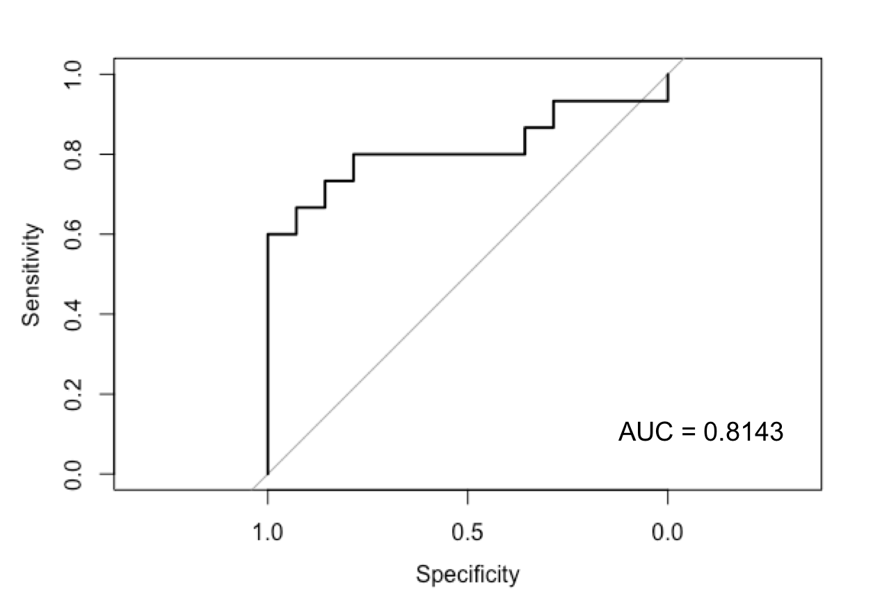
Figure 1: The Receiver Operating Curve (ROC) for
the Random Forest Prediction Model classifying sex using 5 factors: tNAA/tCr,
GSH/tCr,
tCho/tCr,
Glx/tCr
and GM proportion of tissue within the voxel.
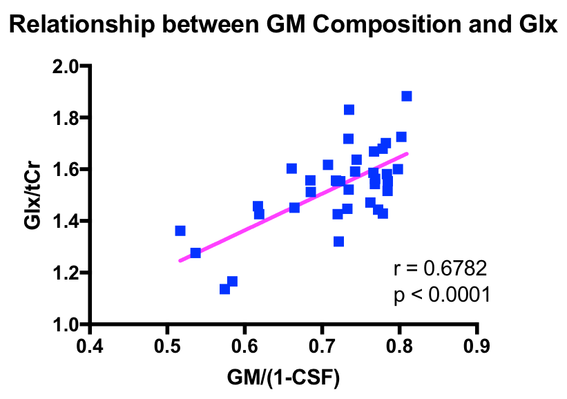
Figure 2: A strong positive correlation between GM
proportion of tissue in the voxel and Glx/tCr
is observed.
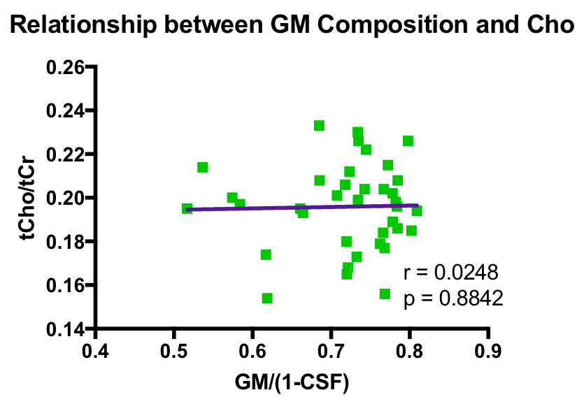
Figure 3: No relationship between GM proportion of
tissue in the voxel and tCho/tCr
is observed.
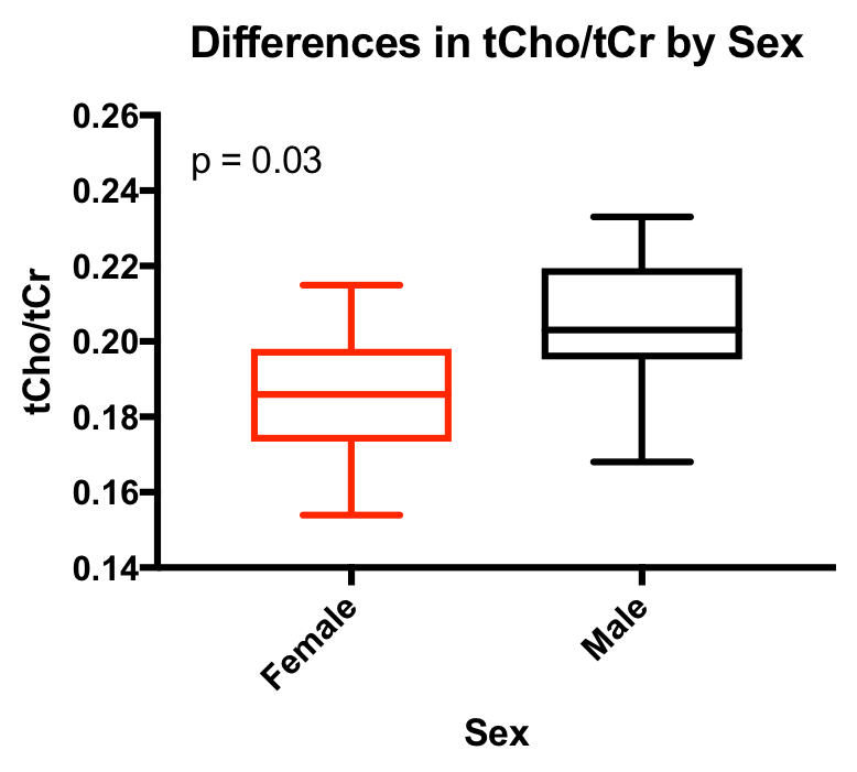
Figure 4: Significantly different tCho/tCr
is observed between males and females. Females in this analysis have lower
concentrations of tCho/tCr.