2266
Impact of Cannula Implantation on Neurochemical Profile in juvenile rat model for ADHD @ 11.7T1Core Facility Small Animal Imaging, University Ulm, Ulm, Germany, 2Center for Magnetic Resonance Research, University of Minnesota, Minneapolis, MN, United States, 3Institute of Anatomy and Cell Biology, Ulm University, Ulm, Germany, 4Core Facility Small Animal Imaging, Ulm University, Ulm, Germany
Synopsis
To detect subtle changes in the neurometabolite concentrations by MRS, it is of utmost importance that the dominant contribution to the metabolic changes, is caused by the pathology of interest itself. Many studies involve surgery to interface e.g. cannulas to the brain. We investigate the impact of implanting an intracerebral cannula on the striatal neurochemical profile. MRS of the striatum from both hemisphere was performed 14d after surgery. A significant reduction of almost all metabolites was observed in the hemisphere with cannulation as well as contralateral side, indicating a dominant impact of the surgery, which might impact the sensitivity of MRS for quantification of pathologic processes.
Purpose:
Intracerebral cannula implantation is a widely used technique for site specific administration of neuroactive substances, pharmacological agents and neuronal tracers, etc., in neuropharmacological and neurological studies, including region-specific brain function (1,2). This can also be employed to implant a microdialysis probe to allow sampling of extracellular fluid in the brain for measuring neurotransmitter concentrations (3). However, this procedure may cause parenchymal damage and consequently inflammatory reaction (4). In this study, a dedicated optimized STEAM sequence with single-shot phase and frequency correction, and image-based shimming was applied to investigate differences in the metabolic profile of the bilateral striatum after unilateral saline microinjections in the dorsolateral striatum in juvenile spontaneously hypertensive rats (SHR) rats with those of similar age in unoperated animals as a control group, using in vivo proton magnetic resonance spectroscopy (1H-MRS) at 11.7T.Methods:
Spontaneously hypertensive rats (SHR) underwent cannula implantation at postnatal day 28 and unilateral intrastriatal injection of saline “Sham_op_inj” at postnatal 42 (n=6). Control group are animals which did not undergo surgery (n=10). A home-built head restrainer was used to properly immobilize the animal's head during measurements, ensuring stability and reproducibility of the experimental setup. Experiments were performed at a dedicated small animal system (117/16 USR BioSpec, AVANCE III, ParaVision 6.01, Bruker BioSpin, Ettlingen, Germany) equipped with a 9 cm inner diameter self-shielded gradient coil insert providing 750 mT/m maximal strength in 80 μs rise time. A 72 mm birdcage quadrature volume resonator was used for excitation and a receive-only rat brain 2x2 element surface coil array was used for signal reception. Volume-of-interests (VOI) were planned based on T1-weighted 2D FLASH (TR/TE = 193/5ms, flip angle 17.5°) images. Field homogeneity was adjusted individually for each investigated region using a field-map based approach (MAPSHIM). A short-echo-time STEAM spectroscopy sequence (TR/TE/TM: 5000/3.5/10ms, 256 acquisitions) combined with VAPOR water suppression was used (5,6). In vivo 1H MR spectra were acquired from 7.9-15.2μl striatum volume located in vicinity of the injection side as well as contralateral side on postnatal day (PND) 42. Spectra were also acquired from 18.5μl striatum volume in control animals on the same age (PND 42). Single-shot data were frequency and phase corrected prior to summation (7). Unsuppressed water signal was used as an internal reference as well as for eddy current correction and absolute metabolite concentrations were derived with LCModel (spectrum fitted from 0.5–4.2 ppm) (8). Statistical significance of the differences was analyzed with the non-parametric Mann–Whitney U-test.Results and Discussion:
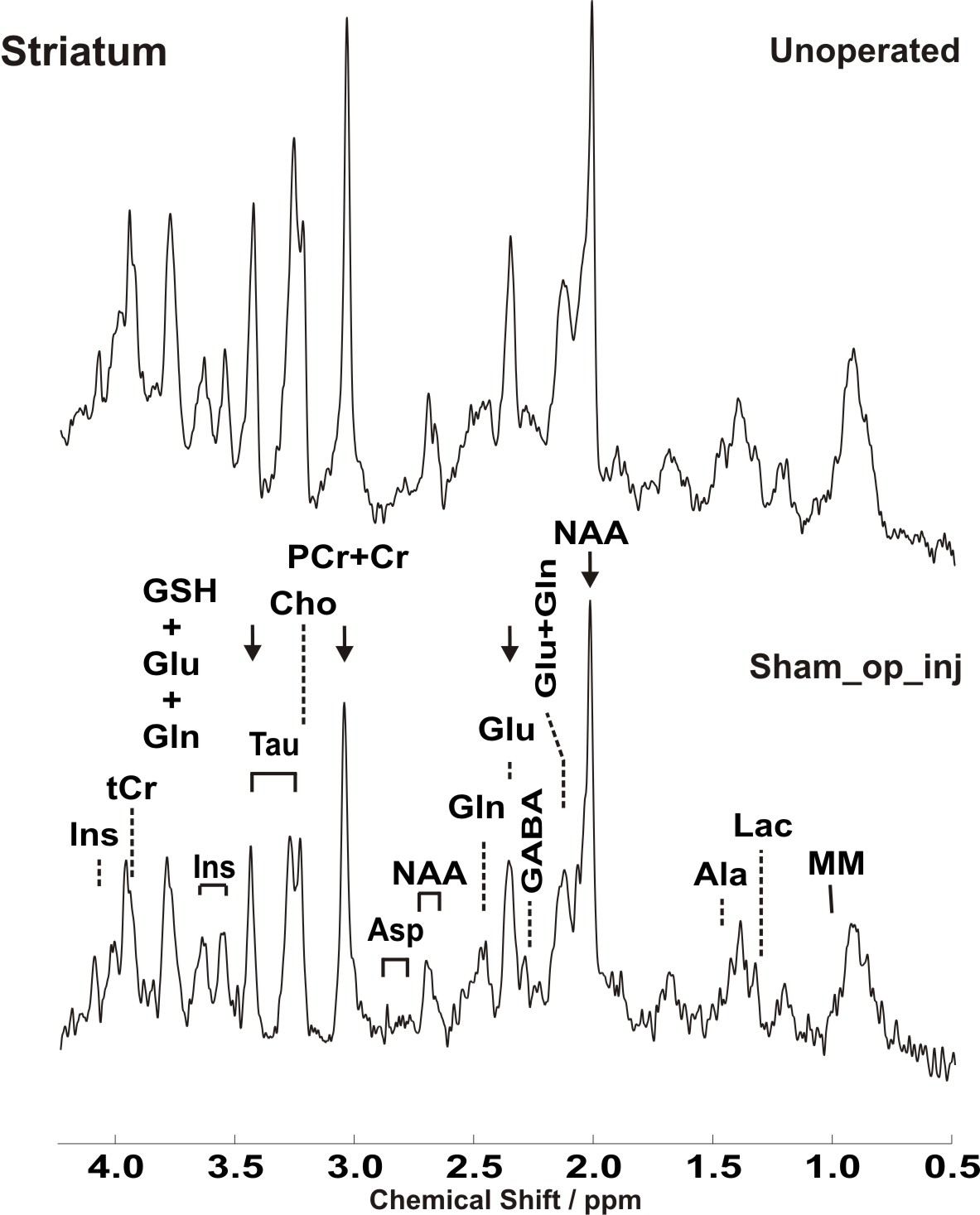
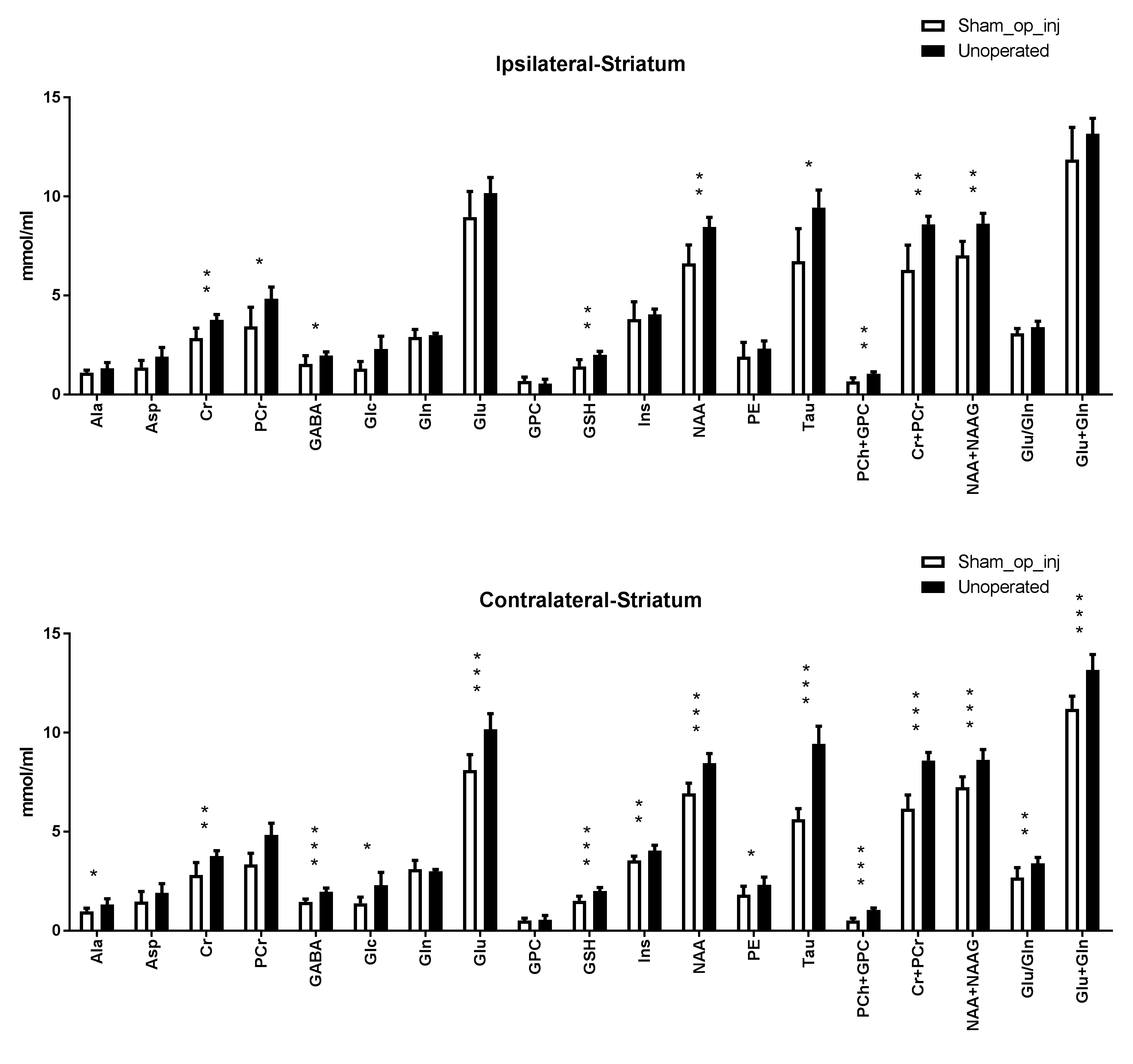
Representative water-suppressed in vivo proton MR spectra of the investigated striatal regions clearly show obvious changes in the neurochemicals concentration (Fig. 1, (9)) for Sham_op_inj group compared to control group. The average full width at half-maximum found by LCModel was 0.026 ± 0.002 ppm (13.0 ± 1.0 Hz) in the striatum. Corresponding SNR were 14.6 ± 3. The low average CRLBs of 8, 3 and 6 for GABA, Glu and Gln in the striatum, proves the reliability of the quantification of the metabolites of the glutamatergic and GABAergic neurotransmission systems. The high spectral quality achieved over the entire chemical shift range (0.5–4.2 ppm) ensured reliable and reproducible quantification of each of the brain metabolites. A detailed comparison of striatal neurochemical profiles of Sham_op_inj with unoperated animals is shown in Fig. 2. Cannula implantation along with saline intracerebral microinjection, led to significant reduction in most of the neurochemicals measured in the bilateral striatum. Despite of the fact that scyllo-Ins and N-acetylaspartylglutamate (NAAG) signal were incorporated into the basis set of LCModel as a model component, corresponding quantification was not reliably possible.Conclusion:
Due to the invasive nature of the surgery, striatal neurochemical profile were still severely altered even after 14 days post cannula implantation. This may hamper the analysis of striatal microinjection effects of neuroactive compounds. In line with previous observation in TBI (10), these data show the sharp alteration of neurochemicals in response to surgery, caused by numerous pathological cellular processes, including inflammation, oxidative stress, mitochondrial dysfunction, excitotoxicity, edema, and hypoxia, as result of brain tissue damage. An optimized short TE STEAM sequences in combination with advanced single-shot frequency and phase correction, and image-based shimming enables the quantification of brain metabolites with high spectral fidelity and reproducibility, demonstrating neurochemical profile alteration of the unilateral intrastriatal saline injected animals in comparison to unoperated animals at very high magnetic field (11.7 T).Acknowledgements
The authors would like to acknowledge the following funding sources: P41 EB015894.References
1. Packard K, Pohorecky LA, Brick J. A simple cannula for intraventricular drug administration in rodents. J Neurosci Methods. 1984 Feb;10(2):139-43.
2. Kokare DM, Shelkar GP, Borkar CD, Nakhate KT, Subhedar NK. A simple and inexpensive method to fabricate a cannula system for intracranial injections in rats and mice. J Pharmacol Toxicol Methods. 2011 Nov-Dec;64(3):246-50.
3. Ellenbroek B, Youn J. Rodent models in neuroscience research: is it a rat race? Dis Model Mech. 2016 Oct 1;9(10):1079-1087.
4. Williams LR, Vahlsing HL, Lindamood T, Varon S, Gage FH, Manthorpe M. A small-gauge cannula device for continuous infusion of exogenous agents into the brain. Exp Neurol. 1987 Mar;95(3):743-54.
5. Tkac I, Starcuk Z, Choi IY, Gruetter R. In vivo 1H NMR spectroscopy of rat brain at 1 ms echo time. Magn Reson Med (1999) 41:649.
6. Abaei A, Rizzo F, Deelchand D, Subgang A, Schneider J, Ludolph A, Rasche V. Region-specific Neurochemical profile differences in juvenile rat model for ADHD and control strain: a 1H MRS study @ 11.7T. In: Proc 24th Scientific Meeting ISMRM; 2016:2407.
7. http://www.cmrr.umn.edu/downloads/mrspa
8. Provencher, Automatic quantitation of localized in vivo 1H spectra with LCModel. NMR Biomed (2001) 14:260.
9. Rizzo F, Nespoli E, Abaei A, Bar-Gad I, Deelchand DK, Fegert J, Rasche V, Hengerer B, Boeckers TM. Aripiprazole Selectively Reduces Motor Tics in a Young Animal Model for Tourette's Syndrome and Comorbid Attention Deficit and Hyperactivity Disorder. Front Neurol. 2018 Feb 13;9:59
10. Janna L Harris, Hung-Wen Yeh, In-Young Choi, Phil Lee, Nancy E Berman, Russell H Swerdlow, Sorin C Craciunas, and William M Brooks. Altered neurochemical profile after traumatic brain injury: 1H-MRS biomarkers of pathological mechanisms. J Cereb Blood Flow Metab. 2012 Dec; 32(12): 2122–2134.
Figures