2264
Feasibility of Single Voxel Proton Spectroscopy of Pineal Gland at 7T1Radiology, University of Pennsylvania, Philadelphia, PA, United States, 2Psychiatry, University of Colorado, Aurora, CO, United States
Synopsis
Taurine is an important metabolite present in the pineal gland and is involved in the regulation of melatonin, which is plays a key role in regulation of sleep-wake cycle, in endocrine metabolism and in depression. However, there have been no known in vivo studies that measured neuro-metabolites in the pineal gland. In this study, for the first time, we explored the feasibility of single voxel proton spectroscopy from pineal gland in vivo in healthy human volunteers at 7.0T MRI.
Introduction
Human pineal gland is a
pea-sized gland located in the middle of the brain between two hemispheres, where
the thalamus from two lobes join and is also known as the third eye (1). It is
the major site for the indoleamine metabolism with the melatonin hormone
primarily synthesized in this gland (2-8). Melatonin hormone is mainly involved
in the sleep-wake regulation cycle also known as circadian rhythm, as well as
in seasonal cycle (9-11). Pineal gland also regulates some of the endocrine
hormones (apart from melatonin) involved in the sexual activity and also reproductive
system in women (12,13). Biochemical analyses from 15 human pineal glands obtained
post-mortem from patients aged 45-89 years old reveals a high concentration of
taurine in this gland, which is also involved in the regulation of melatonin
synthesis (14,15). However, spectroscopy studies localized on pineal gland in vivo in humans are limited owing due
to its smaller anatomy (5-9 mm in length, 1-5mm in width and 3-5 mm in
thickness) and signal to noise issues at low field strength MRI scanners. We
explored the feasibility of single voxel spectroscopy localized on pineal gland
at 7.0T using 32 channel RF coil and the preliminary results are presented in
this study.Methods
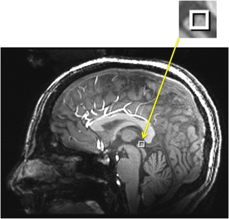
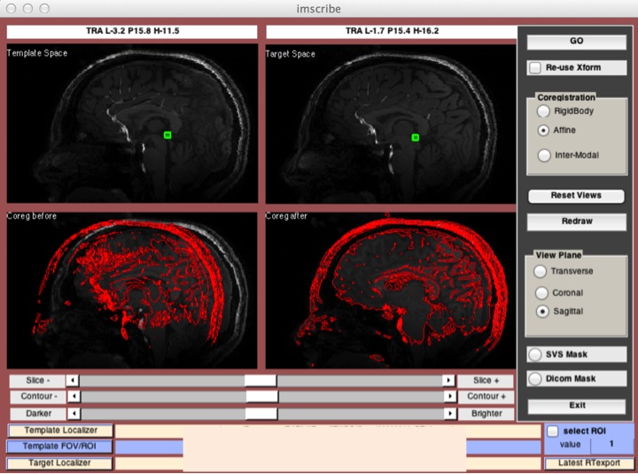
Three healthy volunteers aged 25(F), 38(M) and 68(M) years, participated in this IRB approved study. Single voxel proton spectroscopy of pineal gland (as shown in Figure 1) was acquired on each volunteer post noon using a 7.0T Siemens scanner with a 32-Channel phased-array head coil. In addition one of the volunteer had two additional spectroscopy scans from pineal gland acquired (one at 10am and the other at 6pm) to check the variability in taurine concentration based on the time of the scan. To get the identical spectroscopy voxel for the volunteer for the latter two scans, imscribe software (16) was used as shown in Figure 2. For 1H MRS, the parameters were: voxel size:5x5x5 mm3; TR:3000ms; TE:23ms; dummy scans:4; number of averages for water reference/water suppressed spectra:8/512. Total acquisition time for the spectroscopy was ~27min. Spectra obtained were processed using the LC Model software (17).Results & Discussions
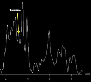
Representative spectrum from pineal gland of the volunteer is shown in Figure 3 with taurine peak represented at ~3.4 ppm. Mean absolute concentration of taurine from the LC model processed spectra was 3.74 +/- 1.2 mM (range 2.52-4.91 mM). For one of the volunteer the taurine concentrations obtained in the morning, post noon and in the evening (10:00am; 2.00pm and 6.00pm) were 3.91, 3.78 and 4.78 mM, respectively. While these preliminary results show the feasibility of single voxel spectroscopy on pineal gland, additional data from more volunteers and also the reproducibility of the measurements of taurine at the same time of the day needs to be established.Summary
This preliminary study demonstrates for the first time the feasibility of singe voxel spectroscopy localized on pineal gland.Acknowledgements
This project was supported by the National Institute of Biomedical Imaging and Bioengineering of the National Institute of Health under award number p41EB015893 and the National Institute of Drug Abuse of the National Institutes of Health under award number R01DA037289.References
1. Macchi MM, Bruce JN. (2004) Human pineal physiology and functional significance of melatonin. Frontiers in Neuroendocrinology 25, 177-195.
2. Lerner AB, Case JD, Takashi Y, Lee TH, Mori N. (1958) Isolation of melatonin, Pineal factor that lightens melanocytes. Journal of American Chemical Society 80, 2587.
3. Lerner AB, Case JD, Heinzelmann RV. (1959) Structure of melatonin. Journal of American Chemical Society 81, 6084-6085.
4. Axelrod J. (1974) The pineal gland: a neurochemical transducer. Science 1184, 1341-1348.
5. Reiter RJ. (1981) The mammalian pineal gland: structure and function. American Journal of Anatomy 162, 287-313.
6. Klein DC. (1985) Photoneural regulation of the mammalian pineal gland, in: Evered D, Clark S (Eds.), Photoperiodism, Melatonin and the Pineal, Ciba Foundation Symposium. Vol. 117, Pitman, London, 38-56.
7. Reiter RJ. (1991) Pineal melatonin: cell biology of its synthesis and of its physiological interactions. Endocrine Reviews 12, 151-180.
8. Klein DC, Coon SL, Roseboom PH, Weller JL, Bernard M, Gastel JA, Zatz M, Iubone M, Rodriguez IR, Begay V, Falcon J, Cahill G, Cassone VM, Baler R. (1997) The melatonin rhythm generating enzyme: molecular regulation of serotonin N-acetyl-transferase in the pineal gland. Recent Progress in Hormone Resesrach 52, 307-358.
9. Lewy AJ, Ahmed S, Jackson JML, Sack RL. (1992) Melatonin shifts human circadian rhythms according to a phase-response curve. Chronobiology International 9, 380-392.
10. Arendt J. (1995) Melatonin and the mammalian pineal gland. Chapman & Hall, London.
11. Arendt J, Skene DJ, Middleton B, Lockley SW, Deacon S. (1997) Efficacy of melatonin treatment in jet lag, shift work, and blindness. Journal of Biological Rhythms 12, 604-617.
12. Binkley S. (1988) The pineal: Endocrine and nonendocrine function. Prentice Hall, Englewood Cliffs, NJ.
13. Kappers JA. (1979) Short history of pineal discovery and research, in: The pineal gland of vertebrates including man, in: Kappers JE, Pevet P. (Eds.). (Elsevier, Amsterdam) Progress in Brain Research 52, 3-22.
14. Vellan EJ, Gjessing LR, Stalsberg H. (1970) Free amino acids in the pineal and pituitary glands of human brain. Journal of Neurochemistry 17, 699-701.
15. Wheler GHT, Klein DC. (1980) Taurine release from the pineal gland is stimulated via a beta-adrenergic mechanism. Brain Research 187, 155-164.
16. Wolf DH, Satterhwaite TD, Loughead J, Pinkham A, Overton E, Elliott MA, Dent GW, Smith MA, Gur RC, Gur RE. (2011) Amygdala abnormalities in first-degree relatives of individuals with schizophrenia unmasked by benzodiazepine challenge. Psychopharmacology 218, 503-512.
17. Provencher SW. (1993) Estimation of metabolite concentrations from localized in vivo proton NMR spectra. Magnetic Resonance in Medicine 30, 672-679.
Figures