2263
Indirect 1H-[13C] NMR Spectroscopy in Rodent Alzheimer’s Model1Integrative Program in Neuroscience, McGill University, Montreal, QC, Canada, 2Brain Imaging Center, Douglas Mental Health Institute, Montreal, QC, Canada, 3Department of Psychiatry, McGill University, Montreal, QC, Canada, 4Comparative Medicine & Animal Resources Centre, McGill University, Montreal, QC, Canada, 5Yale University, New Haven, CT, United States
Synopsis
Alzheimer’s disease (AD) is a neurodegenerative disorder commonly associated with neuronal metabolic deficits. In this study, we employ an indirect 1H-[13C] MRS approach to investigate changes in glucose metabolism between wild type and transgenic Alzheimer rats. This method can be used to follow the labeling of downstream metabolites after [1-6,13C2] glucose infusion and allows for quantitative measurements of the rate of 13C-label transfer from glucose into glutamate and glutamine in the brain. Preliminary comparison of 13C fractional enrichment time courses suggest that transgenic rats exhibit slower 13C metabolite labeling, indicative of a lowered glucose metabolic rate in AD pathology.
Introduction
An estimated 50 million people worldwide suffer from Alzheimer’s Disease (AD), a neurodegenerative disorder characterized by memory loss and progressive cognitive impairment. Despite being the focus of enormous research efforts, there remains a lack of accurate pre-mortem diagnosis and effective treatments against AD. MRS combined with infusion of 13C-labeled substrate is a powerful non-invasive technique that enables investigating brain metabolism in vivo1. This is achieved by infusing a 13C-labelled substrate such as glucose, and subsequently detecting the uptake of 13C-label into downstream metabolic products. Since the 13C-labelled substrates used in MRS are metabolized normally by the brain, 13C-MRS enables a direct measure of the rate of conversion of glucose into downstream metabolic products such as glutamate and glutamine. Given that AD is associated with metabolic deficits, 13C-MRS may be a valuable tool in AD research2.
The aim of this study was to apply 1H and 13C-MRS to detect neurochemical and metabolic alterations in the brain in a rat model of Alzheimer’s disease and to develop quantitative modelling methods for reproducible assessment of neurochemical and metabolic rates in the rat brain. Specifically, the TgF344-AD rodent model3, which exhibits both amyloid and tau pathology as well as neurodegeneration mirroring human AD pathology, was studied. We hypothesized that TgF344-AD rats will exhibit reduced TCA cycle and glutamate/glutamine neurotransmitter cycling rates relative to wild type rats.
Methods
All experimental procedures involving animals were approved by the McGill University Animal Care Committee. Animals were fasted overnight prior to femoral artery and vein cannulation for intravenous infusion of 13C-labeled glucose and periodic blood sampling for further high-resolution NMR analyses of 13C enrichment of substrate. After adjustment of MRS parameters, an exponentially decaying 8-min bolus of 99%-enriched [1,6-13C2] glucose (1.1 M) was administered, followed by a constant infusion over two hours.
Scans were performed on a 7T Bruker Biospec 70/30 horizontal bore preclinical scanner. All MR data were acquired with an in-house 1H-[13C] coil4. An anatomical T1-weighted image was taken using the RARE sequence, TR/TE = 2713/10.8 ms, with a 2-minute acquisition time. A voxel measuring 4x5x5 mm3 was placed in the hippocampus for MRS acquisition (Figure 1). Localized water-suppressed 1H spectra were acquired using the POCE PRESS sequence, TR/TE = 4000/8.13 ms, 6000-Hz spectral width, 128 averages, with an 8 min 48s acquisition time5. One baseline scan was performed in the VOI prior to an infusion of [1,6-13C2] glucose, followed by 15 sequential acquisitions during and following the infusion. Phase correction, frequency correction and averaging of spectra were performed in MATLAB using the FID-A toolkit6. LCModel was used for analyzing the spectra using two in-house simulated basis sets for standard 1H metabolites, and selected 13C enriched metabolites.
A two-compartment neuronal–glial metabolic model was used for analyzing the data from wild type rat7, 8. The TCA cycle rate in the neuronal compartment (VTCA), flux through neuronal glutaminase (Vnt), and Glutamate-alpha–ketoglutarate rate (Vx) were estimated from the fit of data to the metabolic model and other fluxes were fixed to the previously published values9, 10.
Results
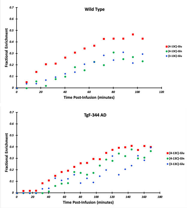
Figure 2 shows an example 1H-MRS time series throughout the two-hour scan duration. A gradual increase of 13C-labelled proton signal for Glu-C4 (2.35 ppm) and Gln-C4 (2.45 ppm) as well as Glx-C3 (2.1 ppm) can be seen. LCModel fitting revealed the dynamics of label uptake in both Glu-C4 and Gln-C4, as well as Glu-C3 (Figure 3). A comparison between the 13C fractional enrichments of the two groups show that while wild type rats reach a steady fractional enrichment of 40% one-hour post infusion, the transgenic rat did not reach 40% FE until two hours post infusion. Metabolic modelling of the wild type rat data revealed flux values for VX, VNt, and VTCA, to be 0.43±0.06, 0.08±0.01, 0.32±0.03 (umol/g/min) respectively, in good agreement results from previous literature10 (Figure 4).Discussion
Preliminary results show that this method can be used to follow and quantitatively measure the rate of 13C-label transfer from glucose into glutamate and glutamine in the brain. Preliminary comparison of fractional enrichment time courses between wildtype and transgenic rats seem to suggest that TgF344-AD rats exhibit slower 13C-labeling, which is indicative of a lowered glucose metabolic rate in AD pathology. The estimated cerebral metabolic fluxes in wild type rats were comparable with previously published results. Data collection and modelling of the transgenic cohort is ongoing.Conclusion
The 13C-MRS methods implemented in this study will provide valuable complimentary information to FDG-PET by revealing, in vivo, which specific glucose metabolism pathways are altered in AD and will allow non-invasive detection of pre-symptomatic changes in the brain.Acknowledgements
The authors thank Andrée Gravel and the CUSM drug discovery platform for her help in acquiring high-res NMR data from plasma samples.References
1. de Graaf, R. A., Rothman, D. L., & Behar, K. L. (2011). State of the art direct 13C and indirect 1H-[13C] NMR spectroscopy in vivo. A practical guide. NMR Biomed, 24(8), 958-972. doi:10.1002/nbm.1761
2. Lin, A. P., Shic, F., Enriquez, C., & Ross, B. D. (2003). Reduced glutamate neurotransmission in patients with Alzheimer's disease -- an in vivo (13)C magnetic resonance spectroscopy study. MAGMA, 16(1), 29-42. doi:10.1007/s10334-003-0004-x
3. Cohen, R. (2013). A transgenic Alzheimer rat with plaques, tau pathology, behavioral impairment, oligomeric aβ, ad frank neuronal loss. The Journal of Neuroscience. doi:10.3410/f.717999415.793479862
4. Kumaragamage, C., Madularu, D., Mathieu, A. P., Lupinsky, D., Graaf, R. A., & Near, J. (2018). Minimum echo time PRESS-based proton observed carbon edited (POCE) MRS in rat brain using simultaneous editing and localization pulses. Magnetic Resonance in Medicine, 80(4), 1279-1288. doi:10.1002/mrm.27119
5. Kumaragamage, C., Madularu, D., Mathieu, A. P., Feyter, H. D., Rajah, M. N., & Near, J. (2017). In vivo proton observed carbon edited (POCE) 13C magnetic resonance spectroscopy of the rat brain using a volumetric transmitter and receive-only surface coil on the proton channel. Magnetic Resonance in Medicine, 79(2), 628-635. doi:10.1002/mrm.26751
6. Simpson, R., Devenyi, G. A., Jezzard, P., Hennessy, T. J., & Near, J. (2015). Advanced processing and simulation of MRS data using the FID appliance (FID-A)-An open source, MATLAB-based toolkit. Magnetic Resonance in Medicine, 77(1), 23-33. doi:10.1002/mrm.26091
7. Duarte, J. M., Lanz, B., & Gruetter, R. (2011). Compartmentalized Cerebral Metabolism of [1,6-(13)C]Glucose Determined by in vivo (13)C NMR Spectroscopy at 14.1 T. Frontiers in neuroenergetics, 3, 3. doi:10.3389/fnene.2011.00003
8. Gruetter, R., Seaquist, E. R., & Ugurbil, K. (2001). A mathematical model of compartmentalized neurotransmitter metabolism in the human brain. American Journal of Physiology-Endocrinology and Metabolism, 281(1). doi:10.1152/ajpendo.2001.281.1.e100
9. Henry, P., Adriany, G., Deelchand, D., Gruetter, R., Marjanska, M., Öz, G., . . . Uğurbil, K. (2006). In vivo 13C NMR spectroscopy and metabolic modeling in the brain: A practical perspective. Magnetic Resonance Imaging, 24(4), 527-539. doi:10.1016/j.mri.2006.01.003
10. M., M. D., Lanz, B., Duarte, J. M., Kunz, N., & Gruetter, R. (2016). Refined Analysis of Brain Energy Metabolism Using In Vivo Dynamic Enrichment of 13C Multiplets. ASN Neuro, 8(2), 175909141663234. doi:10.1177/1759091416632342
Figures