2262
Long-term sensory stimulation causes slow GABA rise after fast glutamate rise: E/I balance monitored by 1H-MRS in awake mice brain1Department of Functional Brain Imaging Research, National Institutes for Quantum and Radiological Science and Technology, Chiba, Japan, 2Department of Molecular Imaging and Theranostics, National Institutes for Quantum and Radiological Science and Technology, Chiba, Japan, 3Department of Psychiatry, McGill University, Montreal, QC, Canada, 4Department of Neurology, Keio University School of Medicine, Tokyo, Japan
Synopsis
To clarify different temporal alterations of Glutamate (Glu) and GABA levels for excitatory/inhibitory (E/I) balance during neural activity in awake healthy mice, we performed 1H-MRS during whisker stimulation. The
Introduction
The balance between excitation and inhibition (E/I balance) of neural activity is fundamental in maintaining normal brain function. The neurotransmitters glutamate (Glu) and gamma-aminobutyric acid (GABA) are essential for E/I balance due to their respective excitatory and inhibitory roles. Disruption of E/I balance may be associated with neurological disorders such as Alzheimer’s disease. Despite the importance of E/I balance for normal brain function and dysfunction, no studies to date have clarified the different behaviors of Glu and GABA in relation to brain function in vivo.Aim
To clarify the different temporal alterations of Glu and GABA levels for E/I balance during neural activity in healthy awake mice.
Methods
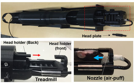
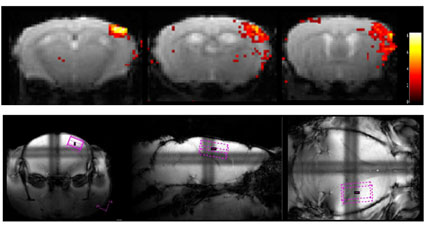
MRI and 1H-MRS MRI and 1H-MRS were performed using a 7.0-T MRI scanner (Bruker Biospin, Germany) with a cryoprobe designed for the murine brain. For awake experiments, we developed a homemade 1H-MRS holder for mice (Fig.1). Mice were anesthetized with 3% isoflurane for <2 min, after which they were placed on the MRI restraint apparatus. After setting the mouse on the holder, the anesthesia was turned off. Magnetic resonance spectra were acquired from a VOI centered in the left barrel cortex (Fig.2). Brain activation was confirmed using functional MRI under whisker stimulation (Fig.2). The size of the voxel was 1.175 mm × 2.0 mm × 3.5 mm. Localized 1H-MRS was applied using PRESS (TE/TR = 11/3000 ms). A total of 292 averages were acquired to obtain sufficient signal-to-noise ratios (e.g., SNR of the NAA peak > 20) for further quantification. Experiments under general anesthesia were performed by inducing anesthesia with 3% isoflurane and maintaining it with 1.5% isoflurane. The remaining manipulations were similar for both awake and anesthetized mice. In vivo spectra were analyzed using the LCModel (Stephen Provencher Inc). Glu and GABA were further analyzed to investigate the alterations of E/I balance during neural activities. Two-photon laser microscopy A cranial window that was 3–4 mm in diameter was attached over the left somatosensory cortex and centered at 1.8 mm caudal and 2.5 mm lateral from the bregma, according to the “Seylaz–Tomita” method (1). The awake mouse was placed on a custom-made apparatus, and real-time imaging was conducted by two-photon laser scanning microscopy (Leica Microsystems GmbH, Germany) with an excitation wavelength of 900 nm. The experimental protocol for two-photon laser scanning imaging measurements was previously reported (2). Statistical analyses Results were presented as the mean ± standard error of the mean. For multiple group comparisons, one-way ANOVA was performed, followed by the Fisher’s least significant difference post-hoc test.Results
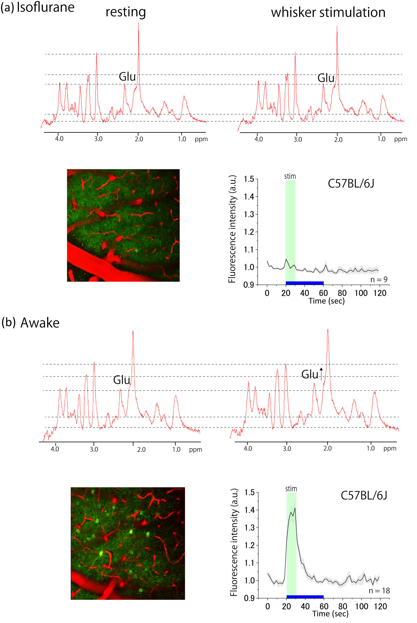
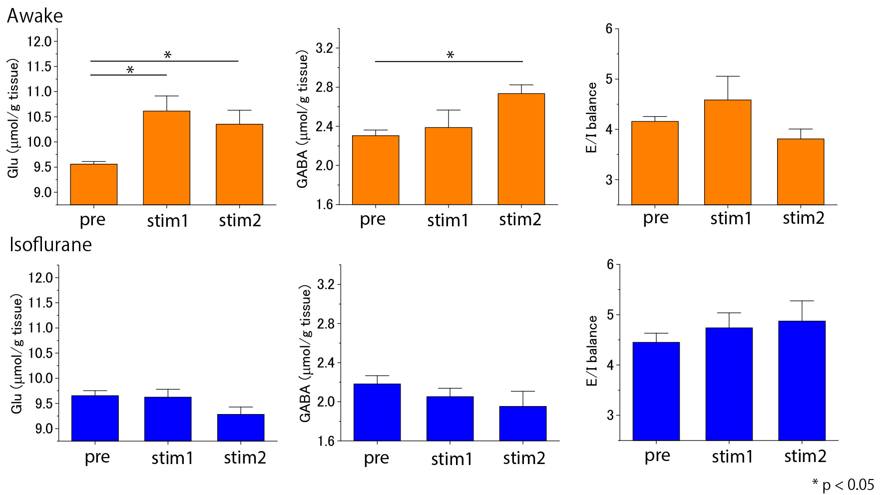
Although no neurochemical alterations were found under anesthesia, there were evident alterations during the awake experiments, which were consistent with the Ca signals imaged by two-photon laser scanning microscopy (Fig.3). In the 1st MRS acquisition with whisker stimulation, a significant increase of the Glu level was observed (p < 0.05) with no significant alteration of the GABA concentration (Fig.4). At the 2nd MRS acquisition with repeated whisker stimulation, the Glu level remained higher than the baseline and GABA also increased significantly compared with the baseline (p<0.05), resulting in a reduced E/I balance (Glu to GABA ratio) (Fig.4).Discussion and Conclusion
Short-term sensory stimulation increased the Glu level, whereas long-term stimulation by repeated whisker stimulation increased GABA levels, leading to a dominantly inhibitory E/I ratio. The decreased E/I ratio may be associated with sensory habituation, which we will confirm in future studies using two-photon laser scanning microscopy. We consider that our experimental protocol with mice may allow observation of habituation based synaptic plasticity, which may be a similar finding in human subjects with overlearning (3). Given the significant influence of anesthesia on neural activity, awake 1H-MRS in mice is a valuable tool to investigate E/I balance in relation to brain function.Acknowledgements
This research is partially supported by the Strategic Research Program for Brain Sciences from Japan Agency for Medical Research and development, AMED.References
1. Tomita et al. J Cereb Blood Flow Metab. 2005;25(7):858-67. 2. Tajima et al. J Cereb Blood Flow Metab. 2014;34(8):1363-72. 3. Shibata et al. Nat Neurosci. 2017;20(10):1427.Figures