2261
Functional MRS of aspartate, glutamate, glutamine and GABA- at 3 Tesla1Emanuel Institute of Biochemical Physics of RAS, Moscow, Russian Federation, 2Radiology, Clinical and Research Institute of Emergency Pediatric Surgery and Traumatology, Moscow, Russian Federation, 3Moscow State University, Moscow, Russian Federation, 4Semenov Institute of Chemical Physics of the Russian Academy of Sciences, Moscow, Russian Federation
Synopsis
In this research visual stimulation was used for the first functional MRS of aspartate, TE averaging – for pure glutamate and glutamine, and MEGA-PRESS for GABA- to find activation-induced changes at 3 Tesla. The results (except for glutamine) are in compliance with previously published data for 7T. The parameters behavior might reflect the manifestation of Glu neurotransmitter function predominance and metabolic character of changes in the concentrations of other parameters studied.
Introduction
The balance of neurotransmitters in response to neuronal activation is one of the key characteristics of central nervous system functioning. The metabolic and neurotransmitter functions of GABA and glutamate (Glu) nowadays are of high interest, so the concentrations of their metabolism participants (aspartate (Asp), NAA) during activation have to be studied. In this research visual stimulation was used for functional MRS of aspartate at 3 Tesla; also, TE averaging1 was used to find the changes of pure Glu and glutamine (Gln) during activation. The stimulation effect on GABA not contaminated with macromolecular fraction (GABA-) was found as well.Materials and Methods
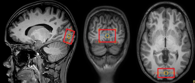
Philips Achieva dStream 3T, SENSE Head-8 coil and InVivo Sensavue (for stimulus transmission) were used. Fifty-seven healthy subjects (age 24.1±3.2) without TBI or any other neurological disorders participated in the study. Each subject passed the standard MRI examination (3D T1w, T2w, FLAIR, DWI). The study consisted of 3 parts, all subjects were divided in three groups: 1) GABA-: 17 participants 2) Glu+Gln: 20 participants 3) Asp: 20 participants The diagnostic study length was ~20 minutes, the fMRS part did not exceed 20 minutes. The 8 Hz flashing checkerboard was used for stimulation; the video was demonstrated through the mirror. Big red crosses were randomly put into the video, the participant had to count these crosses and push the special button when the cross appeared every 20 times. The duration of stimulation was equal to the duration of fMRS data obtaining (~10 mins). Resting spectrum was always performed at first, after that – spectrum during stimulation. The spectroscopy voxel (20x40x30 mm) location in visual cortex (equal in all 3 parts of the study) is demonstrated on fig.1. Shimming and water suppression parameters were the same for rest and stimulus spectra.
For GABA- MEGA-PRESS sequence was used, 20 ms editing pulses, δOn=1.9 ppm, δOff=1.5 ppm, TE=80 ms, TR=2000 ms, NSA=288, (9 min 32 s). For Glu and Gln signals TEavg was used, TE=35, 45, …185 ms, NSA for each TE=16, TR=2000 ms (8 min 32 s).
For Asp signal obtaining: MEGA-PRESS sequence, 27 ms editing pulses, δOn=3.89 ppm, δOff=5.21 ppm2, TE=90 ms, TR=2000 ms, NSA=288, (9 min 32 s). Home-made program based on FID-A3 was written to compile PRESS spectra with TE=90 from OFF-series (PRESS-90). GABA- spectra were processed in Gannet. The result were GABA-/Cr values in each spectrum (further called GABA-). TEavg spectra processing was performed in LCModel. Brain in vivo TEavg spectrum was simulated in FID-A, basis set for LCModel was created, Glu/Cr, Glx/Cr and NAA/Cr values (further called Glu, Glx and NAA) were obtained in each spectrum. Gln was found as Glx-Glu. Asp peak intensity (IAsp) was quantified in AMARES (jMRUI). PRESS-90 spectra were processed in LCModel, INAA, ICr and IGlx were found. For each spectrum all intensities of metabolites were normalized on ICr (IAsp/ICr=Asp, INAA/ICr=NAAPRESS-90, IGlx/ICr=Glx PRESS-90). The relative effect (stimulation divided by rest) was found for GABA-, Asp, Glu, Gln, Glx, and NAA. All relative values were compared with the value=1 (Mann Whitney).
Results
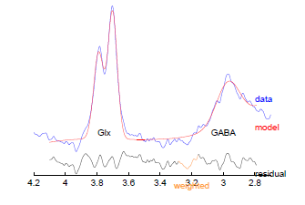
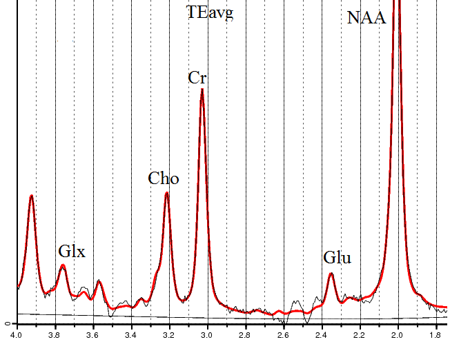
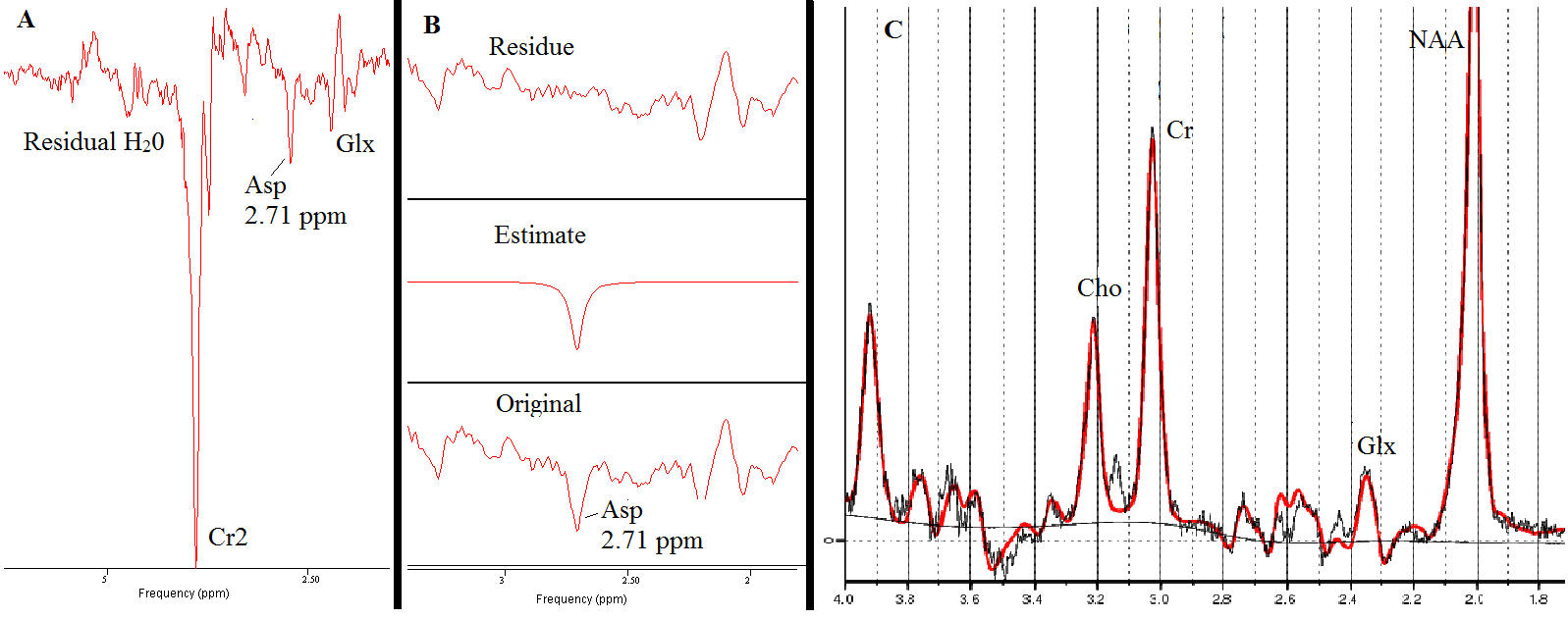
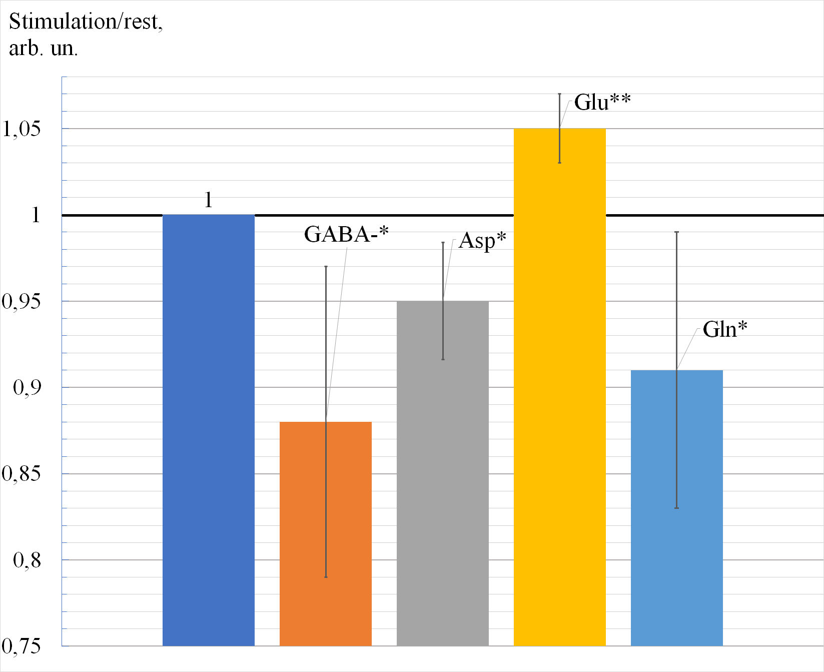
Typical spectra obtained in the study are demonstrated on figures 2-4. In TEavg spectra Glu peak at 2.35 ppm and Glx peak at 3.75 ppm were perfectly fitted by the simulated basis set. The signal of Asp was easily approximated by one Lorentzian curve. Visual stimulation caused the decrease of GABA-, Asp and Gln; Glu was increased, Glx and NAA remained unchanged, see fig. 5.Discussion
To our knowledge, functional MRS of Asp was performed at 3T for the first time. The SNR of Asp peak obtained in the study is enough for the confident quantitative analysis. The decrease in Asp by 5% in response to continuous neuronal activation is in good agreement with previous findings at 7T4, as well as the 12% GABA- reduction5. TEavg allowed the confident observing of the increase in Glu (in agreement with4). We found the decrease in Gln, contraindicative with4.
In this study we tried to characterize the relationship between metabolic and neuromediator functions of the compounds studied during continuous visual activation. The decrease of GABA and Asp could signify the decrease in their synthesis. The Gln decrease might point at the shift in Glu-Gln cycle towards Glu synthesis. All above might be the manifestation of Glu neurotransmitter function predominance and metabolic nature of changes in the concentrations of other parameters studied. Glu pool appears to be maintained by all pathways.
Acknowledgements
This work is supported by the the grant RSF 18-13-00030References
1. R. Hurd et al. Measurement of brain glutamate using TE‐averaged PRESS at 3T. MRM (2004), 51, 3, pp 435-440. https://doi.org/10.1002/mrm.20007
2. P. Menshchikov et al. Quantification of cerebral aspartate concentration in vivo using proton magnetic resonance spectroscopy. Bull. Lebedev Phys. Inst. (2017) 44: 56. https://doi.org/10.3103/S1068335617030022
3. Simpson, R., Devenyi, G. A., Jezzard, P. , Hennessy, T. J. and Near, J. (2017), Advanced processing and simulation of MRS data using the FID appliance (FID‐A)—An open source, MATLAB‐based toolkit. Magn. Reson. Med., 77: 23-33. doi:10.1002/mrm.26091
4. P. Bednařík et al. Neurochemical and BOLD Responses during Neuronal Activation Measured in the Human Visual Cortex at 7 Tesla. JCBFM (2015), Vol 35, Issue 4, pp. 601-610. https://doi.org/10.1038/jcbfm.2014.233
5. C. Chen et al. Activation induced changes in GABA: Functional MRS at 7 T with MEGA-sLASER. NeuroImage (2017) 156, pp. 207-213. https://doi.org/10.1016/j.neuroimage.2017.05.044
Figures