2257
Pharmacologically increased dopamine levels reduce GABA and glutamate concentrations in visual brain areas – a 1H MRS study at 3T1Center for Stroke Research Berlin, Charité – Universitätsmedizin Berlin, Berlin, Germany, 2Department of Psychiatry and Psychotherapy, Charité – Universitätsmedizin Berlin, Berlin, Germany
Synopsis
Psychotic symptoms, such as delusions and hallucinations are frequently observed in schizophrenia. Converging evidence suggests a link to dompaminergic neurotransmission, although the underlying mechanisms are not well understood. In this study, dopamine levels were pharmacologically increased in healthy volunteers to investigate possible neurochemical alterations in the visual cortex measured by single volume 1H MRS using the MEGA-PRESS sequence at 3 T. Reduced GABA and decreased glutamate concentrations were found induced by increased dopamine levels. The former might contribute to the perceptual deficits seen in schizophrenia, while the latter supports the theory of glutamate hypofunction in schizophrenia.
Purpose
Schizophrenia is characterized by psychotic symptoms, such as delusions and hallucinations. Converging evidence suggests a link between psychotic symptoms and a dysfunction in dopaminergic neurotransmission1. However, it is still not well understood how an excess in dopaminergic signaling can lead to delusions and hallucinations. In this context, influential theories frame hallucinations as the tendency to perceive actually irrelevant sensory signals as meaningful2, 3. The psychosis-related tendency to experience meaningful percepts in noise can be experimentally measured with perceptual detection tasks that require participants to distinguish pure noise stimuli from stimuli containing a signal. Accordingly, an increased tendency towards perceiving signals in pure noise stimuli has been related to schizophrenia and associated conditions2-4. One possible role of dopamine in such a framework is that increased dopamine levels might affect visual perception via a modulation of the inhibitory neurotransmitter γ-aminobutric acid (GABA) in the visual cortex. Here, altered GABA levels have already been linked to psychotic conditions and to related perceptual perturbations5, 6.
Thus, the aim of this study was to use L-DOPA to directly modulate the dopamine system of healthy human individuals while they engage in a task that was designed to measure psychosis-like misperception of illusory faces in noise and to investigate the effect of increased dopamine levels on GABA in the visual cortex using 1H MRS. It was expected that L-DOPA as compared to placebo would decrease GABA concentrations in the visual cortex.
Methods
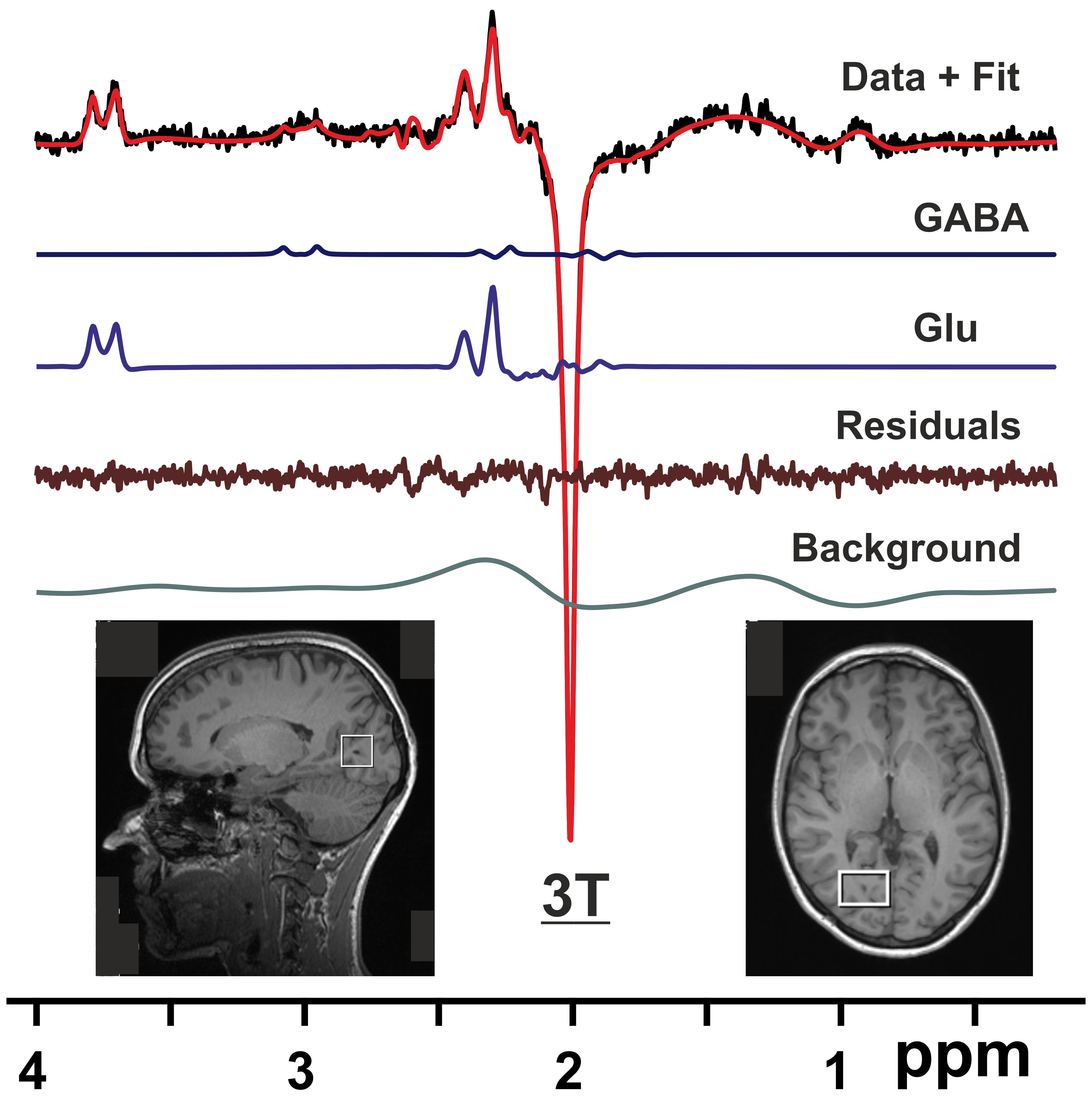
All scans were performed on a 3T Trio system (Siemens Healthineers, Erlangen, Germany) using a 32 channel receive only RF coil. Ten subjects (aged 26 – 48 yrs, 7 f) were scanned twice with a minimum interscan interval of one week (one subject was excluded from MRS analysis due to poor data quality, hence N = 9). One hour prior to each scanning session, subjects were administered orally either 150 mg of L-DOPA or a placebo. All participants and researchers were blinded with respect to the randomly assigned substance administration. For MRS, localized RF calibration was performed, and first- and second-order shims were adjusted using a vendor-supplied algorithm. Voxel placement was performed using a scout scan and high-resolution T1-weighted MPRAGE images. An automatic alignment algorithm was utilized to ensure that the same volume was selected in both sessions. Single volume data from the right visual cortex were acquired using the MEGA-PRESS technique7 with the following scan parameters: VOI = 20x30x20 mm3, TR/TE = 3000/68 ms, number of averages = 128, Tacq = 1024 ms, and editing pulse at 1.9 ppm. MR spectra were pre-processed using the FID-A toolkit8. Metabolite quantification of the resulting difference spectra was performed using LCModel9 with a simulated basis set. Results from MRS were compared for scan sessions “Placebo” and “L-DOPA” using a paired t-test.Results
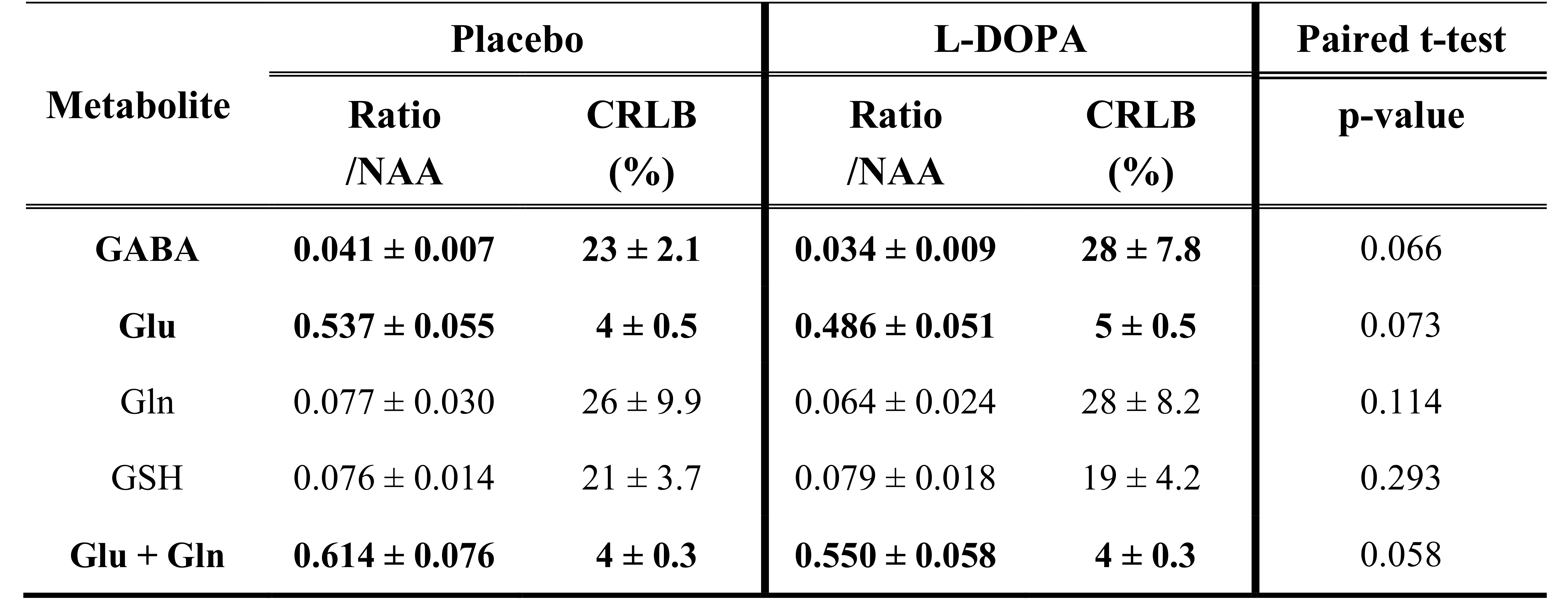
Shimming resulted in water linewidths of 7.3 ± 0.5 Hz across all scans. Sample MEGA-PRESS difference spectra for one volunteer are shown in Fig.1. Results from metabolite quantification (Table 1) show reduced mean ratios of GABA, glutamate (Glu), and combined Glu + glutamine (Gln) with respect to N-acetylaspartate (NAA) for the session “L-DOPA” compared to session “Placebo”. Since it is not expected that the concentration of NAA is altered by the administration of L-DOPA, the reduced ratios translate into reduced concentrations of the aforementioned metabolites. None of the observed differences though reached significance (p<0.05)Discussion
In this study, the dopamine system of healthy volunteers was modulated by the administration of L-DOPA compared to placebo. Using edited 1H MRS it was feasible to observe resulting differences in metabolite ratios of the visual cortex, in particular for neurotransmitters GABA and Glu. That none of the observed differences was significant can partially be attributed to the limited number of subjects included in this ongoing study. However, dopamine-related reduction in GABA levels might contribute to the perceptual deficits (e.g., decreased contrast sensitivity10) seen in schizophrenia and therewith to a diminished sensitivity in distinguishing noise from meaningful information11. Based on the observation that both, antiglutamatergic drugs and antibodies, instantly induce psychosis-like symptoms, a theory of glutamate hypofunction in schizophrenia has been developed12. Further investigation of relationships between increased dopamine levels and decreased glutamate secretion might hence provide insight into the neurochemical alterations underlying positive symptoms in schizophreniaConclusion
Increased dopamine levels due to oral administration of L-DOPA showed a tendency to yield reduced GABA and Glu levels in the visual cortex as assessed by an 1H MRS editing scheme. These findings can contribute to an improved understanding of the mechanism, through which increased dopamine levels cause psychotic symptoms in schizophreniaAcknowledgements
References
1. Howes OD, Nour MM. Dopamine and the aberrant salience hypothesis of schizophrenia. World Psychiatry 2016;15:3-4.
2. Bentall RP, Slade PD. Reality testing and auditory hallucinations: a signal detection analysis. Br J Clin Psychol 1985;24 ( Pt 3):159-169.
3. Hoffman RE, Woods SW, Hawkins KA, et al. Extracting spurious messages from noise and risk of schizophrenia-spectrum disorders in a prodromal population. Br J Psychiatry 2007;191:355-356.
4. Powers AR, Mathys C, Corlett PR. Pavlovian conditioning-induced hallucinations result from overweighting of perceptual priors. Science 2017;357:596-600.
5. Marsman A, Mandl RC, Klomp DW, et al. GABA and glutamate in schizophrenia: a 7 T (1)H-MRS study. Neuroimage Clin 2014;6:398-407.
6. Yoon JH, Maddock RJ, Rokem A, et al. GABA concentration is reduced in visual cortex in schizophrenia and correlates with orientation-specific surround suppression. J Neurosci 2010;30:3777-3781.
7. Mescher M, Merkle H, Kirsch J, Garwood M, Gruetter R. Simultaneous in vivo spectral editing and water suppression. NMR in biomedicine 1998;11:266-272.
8. Simpson R, Devenyi GA, Jezzard P, Hennessy TJ, Near J. Advanced processing and simulation of MRS data using the FID appliance (FID-A)-An open source, MATLAB-based toolkit. Magn Reson Med 2017;77:23-33.
9. Provencher SW. Estimation of metabolite concentrations from localized in vivo proton NMR spectra. Magn Reson Med 1993;30:672-679.
10. Chen Y, Palafox GP, Nakayama K, Levy DL, Matthysse S, Holzman PS. Motion perception in schizophrenia. Arch Gen Psychiatry 1999;56:149-154.
11. Partos TR, Cropper SJ, Rawlings D. You Don't See What I See: Individual Differences in the Perception of Meaning from Visual Stimuli. PLoS One 2016;11:e0150615.
12. Moghaddam B, Javitt D. From revolution to evolution: the glutamate hypothesis of schizophrenia and its implication for treatment. Neuropsychopharmacology 2012;37:4-15.
Figures