2253
1H-NMR based metabolic profiles delineate the anticancer effect of vitamin C on hepatocellular carcinom cell1Department of Electronic Science, Xiamen University, Xiamen, China, 2Johns Hopkins University School of Medicine, Baltimore, MD, United States
Synopsis
Hepatocellular carcinoma (HCC) is a subtype of liver cancer with high worldwide prevalence and mortality. Its high recurrence rate and heterogeneity have challenged effectiveness of existing therapies. Numerous researchers explore the multifaceted benefits of Vitamin C (VC) in cancer treatments. In this study, we utilized the NMR-based metabolomics approach to systematically surrogate the molecular basis underlying the anticancer property of VC in HCC cell. Moreover, combination of VC with chemotherapeutic agent was tried to investigate synergetic effects in modulating metabolic profiles of cancer cells. Our results help to reveal the molecular basis of HCC treatment and may facilitate future clinical therapy designs.
Purpose
Liver cancer is a kind of cancer possessing the third highest mortality worldwide. Particularly, hepatocellular carcinoma (HCC) accounts for the most (70% to 90%) prevalent liver cancers occurring over the world. Characterized by a high recurrence rate and heterogeneity, HCC leads to more than 1 million fatalities annually due to the lack of suitable systemic therapy. Numerous researchers’ ongoing concern is to seek for novel therapeutic modalities towards improved antitumor drugs for advanced or recurrent HCC. As an important natural antioxidant, Vitamin C (VC) is associated with multifaceted benefits to human health. A number of reports have revealed the anticancer potential of high-dose VC. Moreover, broad in vitro and in vivo proofs validate the existence of synergistic effects between VC and other chemotherapeutic agents to benefit cancer treatments. The anticancer property of VC sheds light on the HCC treatment. Pharmacologic VC has been tested on the human HCC cell lines as well as HCC patient-derived xenograft (PDX) models, however, the metabolic basis underlying the anticancer property of VC on HCC remains to be elucidated. Therefore, in this study, we use high-resolution proton nuclear magnetic resonance (1H NMR) -based metabolomics to assess the global metabolic changes in HCC cells induced by VC in combination with oxaliplatin (OXA, chemotherapeutic agent).Methods
Human HCC SMMC-7721 cells were cultured in Dulbecco’s modified Eagle’s medium (DMEM) supplemented with 10% fetal bovine serum at 37°C and 5% CO2. When reached 25% confluency, the cells were randomly divided into four groups for different drug manipulations: treatment with 50 μmol/L oxaliplatin (OXA group, n=10), treatment with 1 mmol/L VC (VC group, n=10), treatment with 50 μmol/L oxaliplatin in combination with 1 mmol/L V C (OXA+VC group, n=10), and without treatment as the standard model control (SM group, n=10). Treatments were administered once for all. Following 48 h incubation, cells were harvested and prepared for NMR experiments. The obtained 1H-NMR spectra datasets were preprocessed and interpreted with multivariate statistical analysis, including principle component analysis (PCA), orthogonal projections to latent structures discriminant analysis (OPLS-DA), Student’s t-test, and metabolite set enrichment analysis (MSEA) to investigate metabolic perturbations in HCC cells after exposure to different treatments. For cell proliferation assay, Cell Counting Kit-8 (CCK-8) assay were also conducted for different groups (OXA, VC, OXA+VC, and SM groups).Results and Discussion
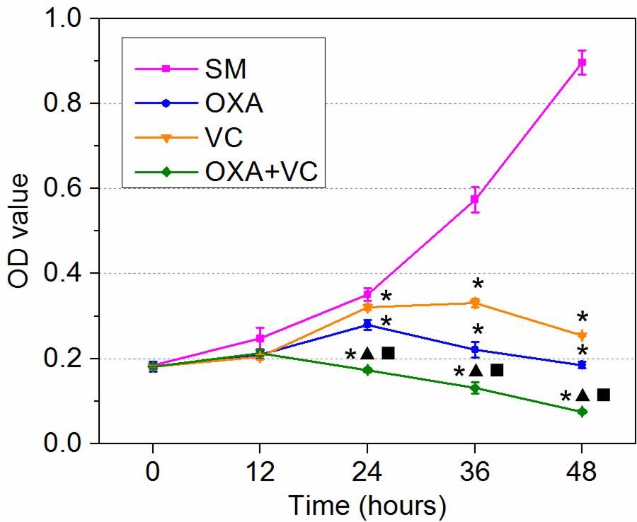
As the CCK-8 assay result shows (Figure1), reductions of proliferation rate in OXA, VC, and OXA+VA groups were observed in comparison with the SM group. Generally, OXA, VC, or OXA+VC treatment can decrease cell proliferation and induce cell death in a time- and drug-dependent manner. Longer treatment duration leads to more prominent inhibition in cell growth. By constrast, OXA+VC treatment possesses the highest treatment efficacy and VC the lowest.
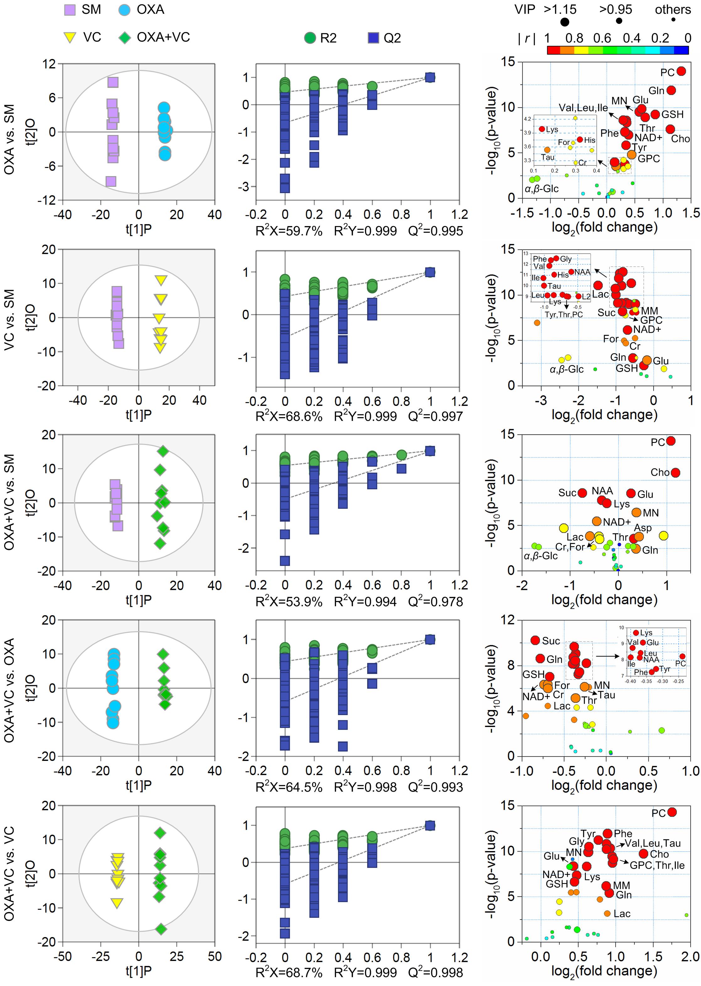
Moreover, the pair-wise OPLS-DA between OXA and SM, between VC and SM, between OXA+VC and SM, between OXA+VC and OXA, and between OXA+VC and VC revealed distinguished group segregations and showed elegant coefficient of determination (R2) as well as good prediction accuracy (Q2) (left and middle panels in Figure 2, respectively). A total of 27 metabolites (a union set of characteristic metabolites from different comparisons) which have primary contributions to group separations were found to be with significant change (p < 0.01) among the four groups (right panel in Figure 2). In comparison with the SM group, most metabolite concentrations in the OXA group have increased; an opposite change has been noticed in the VC group. The OXA and VC group share various common characteristic metabolites with different variation trends. Otherwise, the OXA+VC group exhibits a combination of metabolite change from both OXA and VC groups.
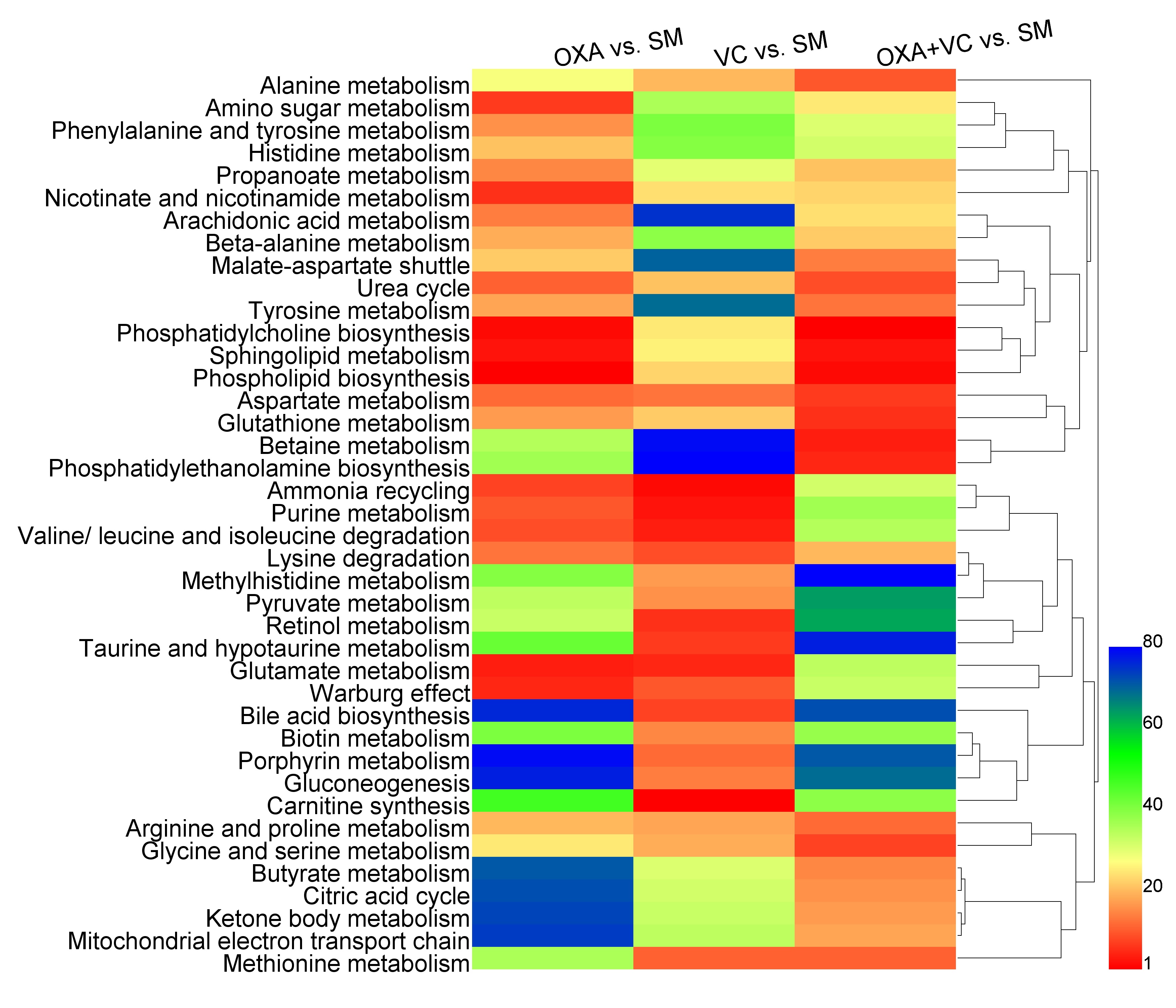
Metabolic pathways with significant variations induced by OXA and VC treatment were revealed by MESA. Figure 3 shows the heat map of clustering analyses on the importance levels of metabolic pathways with significant variations. This heat map provides an additional proof for the conclusion that OXA, VC, and OXA+VC are associated with different mechanism to induce metabolic variations to cells.
Conclusion
According to the NMR-based metabolism analyses, we speculate that OXA and VC are characterized with different mechanisms to modulate the metabolic profile of SMMC-7721 cells. OXA and VC works in unison to induce complementary metabolic modulations, thus resulting in favorable synergistic effects.Acknowledgements
This work was financially supported by the the National Natural Science Foundation of China (No. 81371639). We thank all the members for helpful discussions.References
1. Torre L. A., Bray F., Siegel R. L., et al. Global cancer statistics, 2012. CA-Cancer J. Clin. 2015; 65(2): 87-108.
2. Padayatty S. J., Sun H., Wang Y. H., et al. Vitamin C pharmacokinetics: implications for oral and intravenous use. Ann. Intern. Med. 2004; 140(7): 533-537.
3. Moriguchi M., Umemura A., Itoh Y. Current status and future prospects of chemotherapy for advanced hepatocellular carcinoma. Clin. J. Gastroenterol. 2016; 9(4): 184-190.
Figures