2250
Rapid High-Resolution Metabolic Imaging of Stroke Using SPICE1Institute for Medical Imaging Technology, School of Biomedical Engineering, Shanghai Jiao Tong University, Shanghai, China, 2Radiology department, The Fifth People's Hospital of Shanghai, Fudan University, Shanghai, China, 3Beckman Institute for Advanced Science and Technology, University of Illinois at Urbana-Champaign, Urbana, IL, United States, 4Department of Electrical and Computer Engineering, University of Illinois at Urbana-Champaign, Urbana, IL, United States, 5Shanghai minhang hospital of Integrated traditional Chinese and Western Medicine Hospital, Shanghai, China, 6Department of Biomedical Engineering, Case Western Reserve University, Cleveland, OH, United States
Synopsis
Disrupted metabolic activity is one of the most prominent pathophysiologic consequences of stroke. 1H-MRSI has been recognized as a powerful tool for metabolic imaging, but its clinical applications have been limited due to long scan time and poor spatial resolution. In this study, we investigate the feasibility of rapid high-resolution metabolic imaging of stroke using SPICE (SPectroscopic Imaging by exploiting spatiospectral CorrElation). We have successfully acquired metabolic maps from the whole brain at a nominal spatial resolution of 2.0×2.4×2.0 mm3 in a 5-min scan. Our experimental results clearly show metabolic alterations due to stroke.
Introduction
Imaging markers that can predict brain tissue viability after acute ischemic stroke have been continuously developed over the past four decades1. Despite the promise of diffusion- and perfusion-weighted MRI, there remains a critical need for better biomarkers that can provide more accurate assessment of cerebral pathophysiological conditions of brain tissues. Disrupted metabolic activity is one of the most prominent pathophysiologic consequences of stroke. Metabolic imaging will add significant insights into stroke tissue viability, which could further aid the selection of therapies for patients and assessment of recovery2. MRSI has been recognized as a powerful tool for metabolic imaging, however, most existing MRSI studies in stroke are performed using single-slice MRSI or single-voxel techniques at low resolutions. The long data acquisition time and partial volume effects associated with low-resolution/single-voxel techniques reduce the detection sensitivity. Therefore, despite the potential advantages of whole brain 1H-MRSI, its clinical and research applications in stroke have been extremely limited. In this study, we investigate the feasibility of rapid high-resolution metabolic imaging of stroke using a newly developed 1H-MRSI technology. We have successfully demonstrated that in a 5-min scan, we can acquire metabolic maps from the whole brain at a nominal spatial resolution of 2.0×2.4×2.0 mm3, which clearly show neurometabolic alterations due to stroke.Methods
We used the latest version of the SPICE (SPectroscopic Imaging by exploiting spatiospectral CorrElation) technology3-4 for rapid high-resolution metabolic imaging. The data acquisition scheme has the following novel features: a) FID acquisitions with an ultrashort-TE (1.6 ms) and very-short-TR (160 ms), b) no water/lipid suppression, c) variable density sampling of (k, t)-space, d) rapid and extended k-space coverage with EPSI trajectories, and e) incorporation of navigators for detection and correction of field drift and subject head motion. The data acquisition scheme achieves a nominal resolution of 2.0×2.4×2.0 mm3 in a 5-min scan.
Reconstruction of the spatiospectral function from the SPICE data is accomplished using a union-of-subspaces model, incorporating pre-learned spectral basis functions3-4. These spectral basis functions include the resonance structures of the detectable molecules (generated using quantum mechanical simulations) and their lineshape functions (pre-learned from training data). Spectral quantification is done using an improved LCmodel-based algorithm that incorporates both spatial and spectral priors9.
To help data analysis for our experimental study, we also performed structural imaging scans, which included diffusion-weighted imaging (DWI) (1.3×1.3×4.0 mm3, field of view = 220 mm, b = 0 and b = 1000 s/mm2, TR = 5200 ms, TE = 64 ms, 25 slices), 3D MPRAGE imaging (1.0×1.0×1.0 mm3, field of view = 256 mm, TR = 2500 ms, TE = 2.26 ms, TI=900 ms, 176 slices) and T2-weighted Fluid-Attenuated Inversion Recovery (FLAIR) imaging (0.5×0.5×2.0 mm3, field of view = 240 mm, TR = 9000ms, TE = 89 ms, 82 slices). These structural images helped identify the lesion areas for data analysis. All the scans were performed on a 3.0T Siemens Skyra scanner with IRB approved by the Fifth People’s Hospital of Shanghai, China.
Results
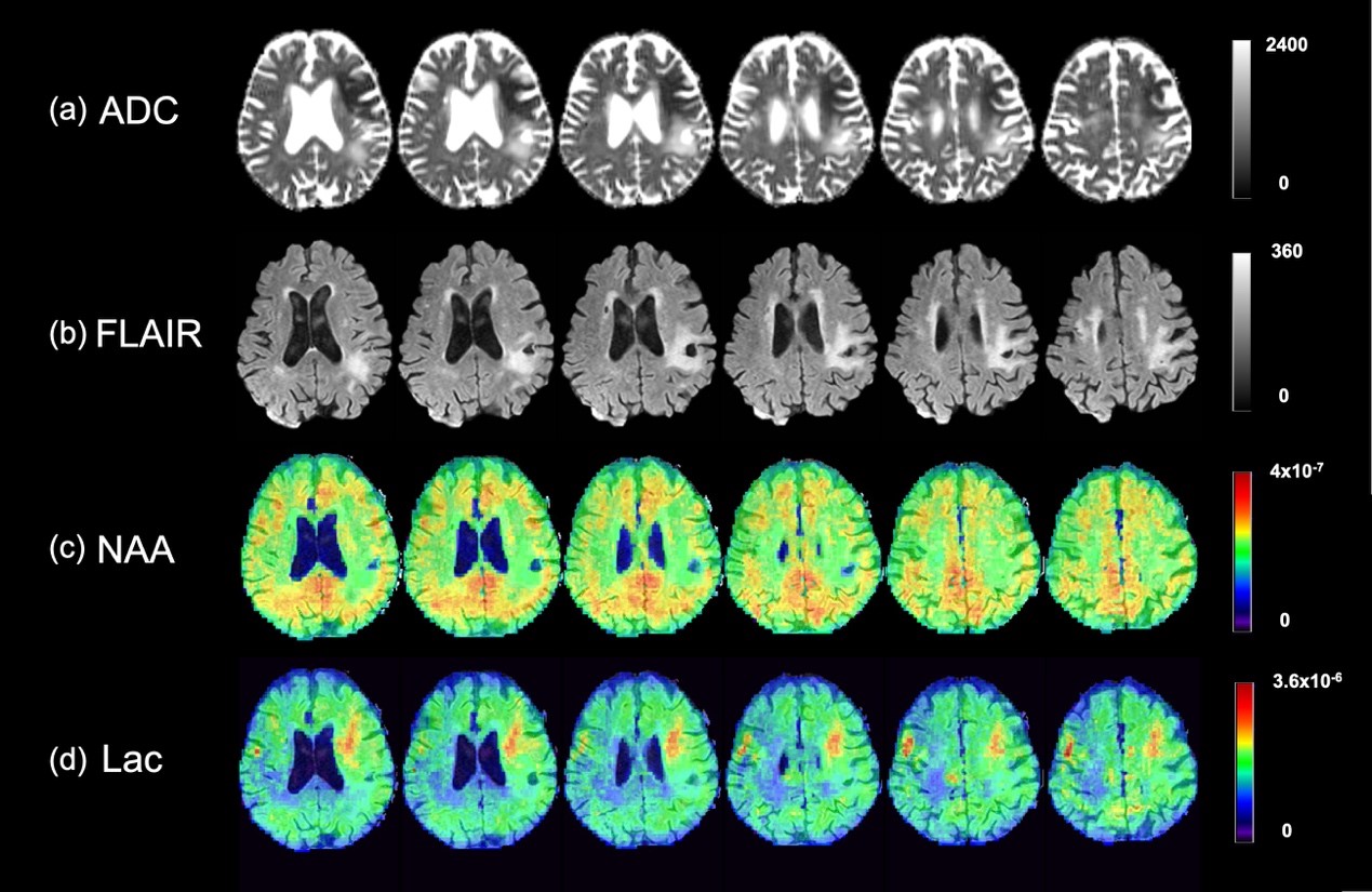
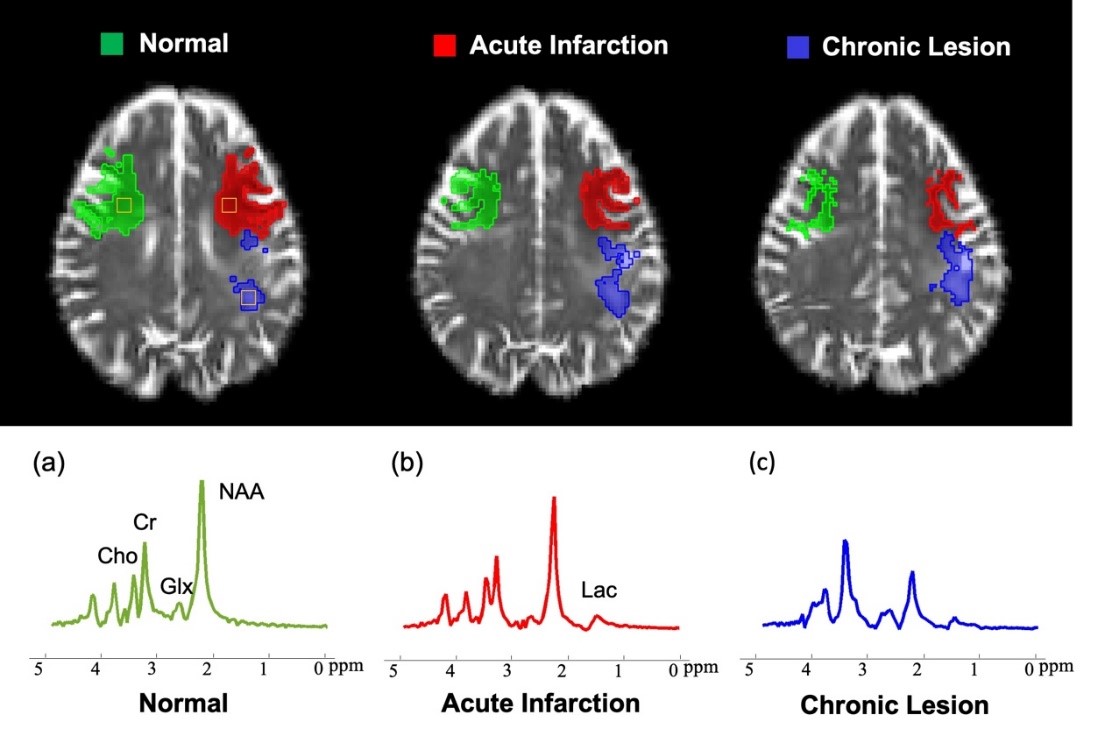
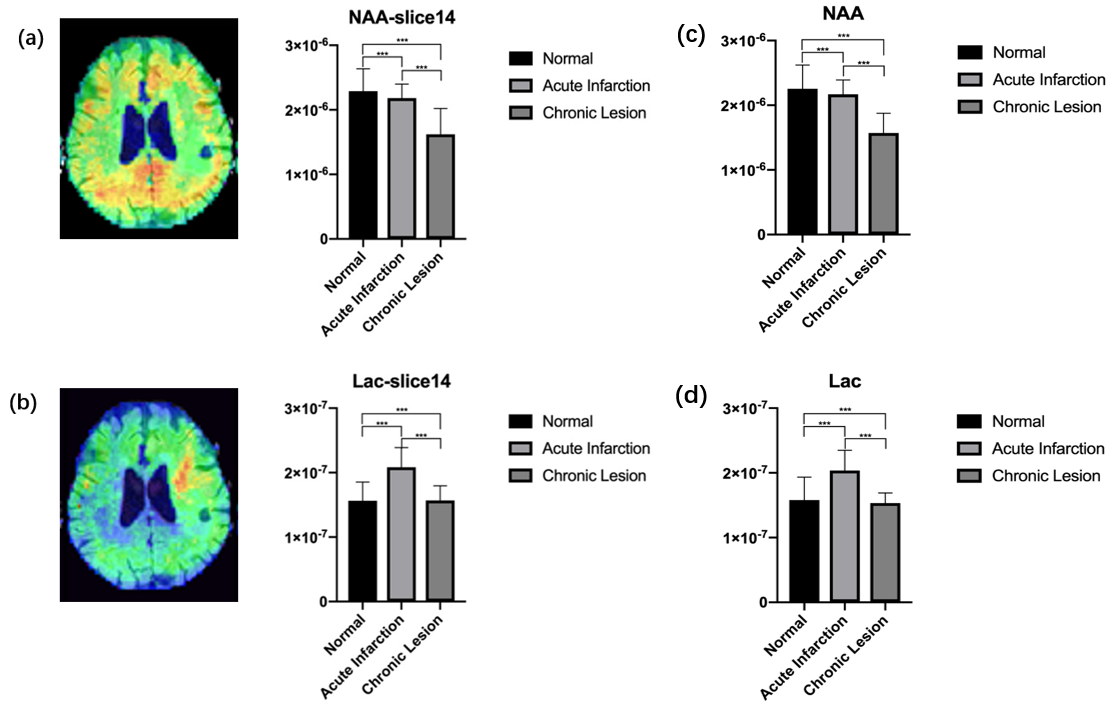
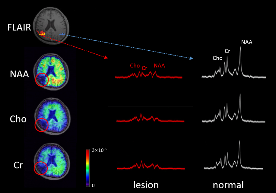
Figure 1 shows the high-resolution NAA and Lac maps of a recurrent ischemic stroke patient with an acute infarction (12h after stroke) and a chronic lesion (3yr after stroke). The corresponding ADC map and the FLAIR map are also included. Figure 2 shows some representative spectra from the acute infarction, chronic lesion and the contralateral normal tissue, respectively. As shown in Fig. 3, the NAA concentration was reduced in both the acute infarction and chronic lesion, and the Lac concentration increased in the acute infarction. Interestingly, the Lac concentration was negatively correlated with the ADC level within the acute infarction, as shown in Fig. 4. This result is consistent with previous findings8. Figure 5 shows another set of results acquired from a more severe stroke patient with a large 5-year chronic lesion. As can be seen, the NAA, Cho and Cr concentrations were all reduced significantly, as expected. Note that these are just some of our representative experimental results; overall, our rapid high-resolution MRSI experiments captured the metabolic alterations due to the strokes very well.Conclusion
This study successfully demonstrates the feasibility of rapid high-resolution metabolic imaging of stroke. In a 5-min scan, we are able to obtain high-quality spatial maps of metabolites at a nominal resolution of 2.0×2.4×2.0 mm3, which clearly capture metabolic changes due to stroke. Our study may lay a foundation for further investigation and application of rapid high-resolution metabolic imaging for accurate assessment of tissue viability in stroke to aid the selection of therapies and to evaluate the therapeutic effects.Acknowledgements
This work is supported by National Science Foundation of China (No.61671292 and 81871083) and Shanghai municipal commission of health and family planning (No. 20154Y0094).References
1. Leigh R, Knutsson L, Zhou J, et al. Imaging the physiological evolution of the ischemic penumbra in acute ischemic stroke. J Cereb Blood Flow Metab 2018, 38:1500-1516.
2. Dani KA, Warach S. Metabolic imaging of ischemic stroke: the present and future, Am J Neuroradiol. 2014, 35:37-43.
3. Fan L, Ma C, Clifford B, et al. High‐resolution 1H‐MRSI of the brain using SPICE: data acquisition and image reconstruction. Magn Reson Med 2016, 76:1059-1070.
4. Peng X, Lam F, Li Y, et al. Simultaneous QSM and metabolic imaging of the brain using SPICE. Magn Reson Med 2018, 79:13-21.
5. Jenkinson M, Beckmann CF, Behrens TE, et al. FSL. Neuroimage 2012, 62:782-790.
6. Harston GW, Okell TW, Sheerin F, et al. Quantification of Serial Cerebral Blood Flow in Acute Stroke Using Arterial Spin Labeling. Stroke 2017, 48:123-130.
7. Ma C, Lam F, Johnson C L, et al. Removal of nuisance signals from limited and sparse 1H MRSI data using a union‐of‐subspaces model. Magn Reson Med 2016, 75:488-497.
8. Cvoro V, Wardlaw JM, Marshall I, et al. Associations between diffusion and perfusion parameters, N-acetyl aspartate, and lactate in acute ischemic stroke. Stroke 2009, 40:767-772.
9. Li Y, Lam F, Clifford B, et al. A Subspace Approach to Spectral Quantification for MR Spectroscopic Imaging. IEEE Trans Biomed Eng 2017, 64:2486-2489.
Figures