2238
Localised 2D J-resolved spectroscopy: Enhanced sensitivity and accelerated acquisition of brain metabolites1Wolfson Brain Imaging Centre, University of Cambridge, UK, CAMBRIDGE, United Kingdom, 2The Department of Clinical Neurosciences, University of Cambridge, Cambridge, United Kingdom
Synopsis
Proton 1D MR spectroscopy has been used to quantify alterations in cerebral metabolite concentrations to investigate neurological diseases. However, the J-coupling found in key metabolites produce multiplets which result in highly overlapped signals, complicating signal quantification. 2D J-resolved spectroscopy (JPRESS) allows distribution of coupled signals into a second perpendicular dimension, reducing signal overlap. Unfortunately, the translation of 2D-JPRESS into a commonly used clinical tool has not been achieved due to limited post-processing strategies and long scan times. Here, a simple processing strategy to significantly enhance signal sensitivity and a technique to accelerate JPRESS by 30% are presented.
Introduction
In vivo two-dimensional (2D) magnetic resonance spectroscopy (MRS) techniques have allowed quantification of additional metabolites when compared with conventional 1D sequences1. Construction of the second or indirect dimension in 2D-JPRESS allows for metabolite signals to be defined by two frequency coordinates helping to better resolve J-coupled metabolite resonances. 2D-JPRESS is however still not widely used in a clinical setting due to long scan times and limited post processing strategies. Firstly, we present a backward linear predictive processing technique2 to enhance the signal to noise ratio (SNR) of 2D JPRESS, with the goal of enabling more accurate metabolite quantification. Secondly, a strategy facilitating a scan time reduction of 30% is achieved, while maintaining signal sensitivity.
Material & Methods
A 2D JPRESS sequences was modified from a standard point resolved spectroscopic sequence (PRESS). An incremental delay (Δt1) was added either side of the second 180o refocusing pulse. An option to accelerate the scan was added to the exam card, exponentially decreasing the TR as a function of the t1 increment3. This results in slight line broadening of the indirect dimension. All scans were performed on a Siemens 3T Skyra MAGNETOM scanner with a 32-channel head coil. Scans were acquired from the GE Braino phantom, containing physiological cerebral metabolite concentrations. Two sequences variants were used: Standard JPRESS with constant TR and accelerated JPRESS with an exponentially decreasing TR. The following parameters were used: TE = 30 ms, TRmax = 2000 ms, TRmin = 70 ms, Δt1 = 2 ms, voxel size 4cm by 4cm by 4cm, f2 Bandwidth = 2000 Hz, Measurements = 100, Averages = 4, water suppression Bandwidth =70 Hz, scan time = 9 mins 14 secs. Without TR variation scan time was 13 mins 20 secs. Four repeat experiments were taken for both sequences giving a mean value and standard deviation for each metabolite signal. Shimming was performed using GRE shim giving a water full width half maximum (FWHM) value of 3.8 - 4.2 Hz for the Braino phantom.
The following outlines the proposed processing pipe line. Complex data in the indirect dimension were shifted by the number of acquired increments, followed by backward linear prediction of the negative evolution period. Both the indirect (f1) and direct (f2) FIDs were multiplied by an unshifted sine bell window apodization function. All spectra were then analysed in magnitude mode. Processing was done in Bruker’s freely available (under an academic licence) Topspin environment. Rectangular volume integrals were constructed to measure the volume of the peaks of interest. For comparison spectra were also acquired with standard 2D post processing: i) zero filling to twice the number of acquired data points (ii) multiplication by an unshifted sine bell function4.
Results
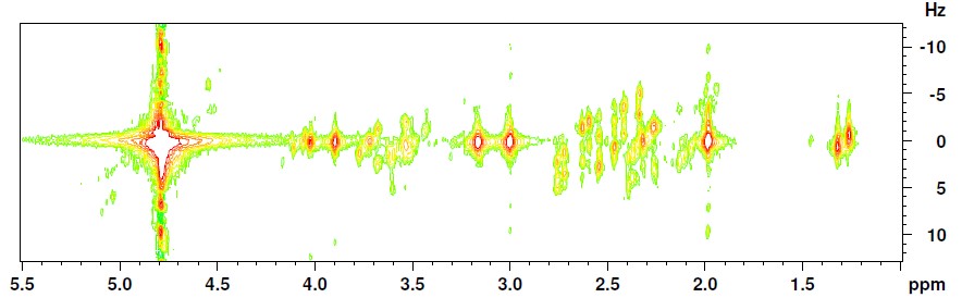
An accelerated JPRESS sequence following the proposed post processing strategy is presented in Figure 1.
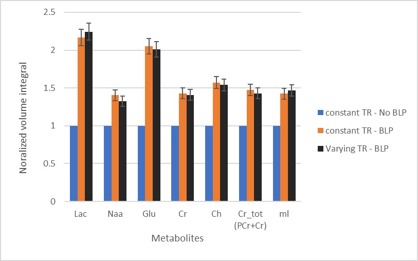
Signal enhancement: Backward linear prediction of the indirectly sampled echo to give a new FID with the shape of a fully sampled echo has been shown to give considerable signal enhancement of key cerebral metabolites in vitro. Figure 2 shows a quantitative representation of this signal enhancement.
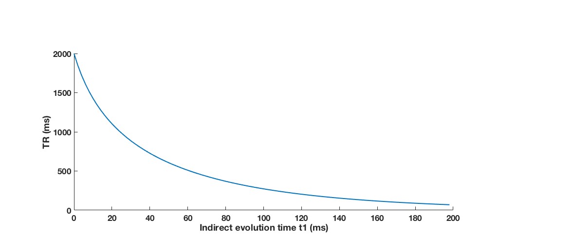
Scan time reduction: Additionally, a reduction (30 %) in scan time was accomplished through varying the repetition time (TR) as a function of the t1 increment (Figure 3). Figure 2 also shows that this accelerated sequence does not influence the quality of signal enhancement.
Discussion
This study outlines a method to improve the SNR of key cerebral metabolites using backward linear prediction of the f1 data of 2D JPRESS. Shifting the f1 data by the number of acquired points allows the resulting data set to match the shape of the sine bell apodization window function, significantly contributing to the reported signal enhancements. Repeat phantom experiments show that these signal enhancements are reproducible. Through TR variation JPRESS could be accelerated while retaining comparable SNR enhancement to the conventional constant TR approach. Critically, as the first data point in the indirect FID is acquired with a long TR, the volume integral of the 2D spectrum still represents the fully relaxed signal3.Conclusion
In conclusion, a new approach to processing in vivo 2D JPRESS data has demonstrated considerable signal enhancement of key brain metabolites. Furthermore, a 30 % scan time reduction is achieved without degrading the quantitative capabilities of our scan. Work in simulating a ProFit1 (2D prior knowledge quantification package) usable basis set that models this backward linear reconstruction is in development.Acknowledgements
Cambridge Trust
Wolfson Brain Imaging Centre
CTR is funded by a Sir Henry Dale Fellowship from the Welcome Trust and the Royal Society [098436/Z/12/B].
References
1. Schulte, R. F. & Boesiger, P. ProFit: two-dimensional prior-knowledge fitting ofJ-resolved spectra. NMR Biomed. 19, 255–263 (2006).
2. Sakhaii, P. & Bermel, W. Improving the sensitivity of conventional spin echo spectra by preservation of initial signal-to-noise ratio. J. Magn. Reson. 242, 220–223 (2014).
3. de Graaf, R. A., De Feyter, H. M. & Rothman, D. L. High-sensitivity, broadband-decoupled 13 C MR spectroscopy in humans at 7T using two-dimensional heteronuclear single-quantum coherence. Magn. Reson. Med. 74, 903–914 (2015).
4. Binesh, N., Yue, K., Fairbanks, L. & Thomas, M. A. Reproducibility of localized 2D correlated MR spectroscopy. Magn. Reson. Med. 48, 942–948 (2002).
Figures