2224
Left Ventricle Segmentation with Densely Connected Full Convolutional Network1Shenzhen Institutes of Advanced Technology, Chinese Academy of Sciences, Shenzhen, China
Synopsis
Cardiac functional analysis is important in heart disease diagnosis. Conventional manual segmentation of left ventricle is time consuming and observer dependent. Our proposed Densely Connected Full Convolutional Network (DenseV-Net) enables automatically process medical images. Its densely connected convolutional block consists of residual calculation with Elu used as active function. The results show that the proposed DenseV-Net can efficiently segment left ventricle from cardiac cines with mean DSC of 0.90±0.12, more accurate compared to V-Net (0.85±0.13, P<0.05). The method offers a feasible way for efficient analysis of cardiac function.
Introduction
Accurate cardiac functional analysis is of importance for diagnosis of heart diseases [1]. To quantitatively evaluate the cardiac function, segmentation of left ventricle is one of the most important steps for left ventricle volume measurement [2]. So far, a number of methods, including semi-automated and automated methods, have been developed for image segmentation [3]. Although semi-automatic methods are easy to be implemented, requirement of extensive manual intervention is time-consuming and observer dependent. With the increasing advancement of deep-learning theory, artificial neural networks emerge as powerful tools in medical image postprocessing. In this study, we proposed an automated LV segmentation method which combines residual function [4] and densely connected full convolutional network (DenseV-Net). Dice coefficient is used to evaluate the performance of proposed method in LV segmentation in a cohort of infarct patients.Materials and methods
Dataset: The study was approved by local ethics committee and informed consent was obtained from each subject. Twenty patients were scanned on a 3T scanner (MAGNETOM TIM Trio, Siemens). Real-time cines were acquired by using balanced steady state free precession imaging with parameters of repetition time (TR) = 2.5 ms, echo time (TE) = 1.1 ms, matrix size = 176×96, fixed field of view (FOV) = 360×270 mm2, in-plane resolution of 2.1×2.8 mm2, and slice thickness = 10 mm with 2 mm slice gap. Slice number was adapted to the heart size so that the whole LV was covered. Parallel imaging with an acceleration rate of 4 using TGRAPPA was used, and the temporal resolution was 61.0 ms. Each slice was imaged for approximate 4.5 seconds to include at least one complete respiratory cycle, which resulted in 73 images. Cines were loaded into ITK-SNAP and segmented manually by an experienced expert.
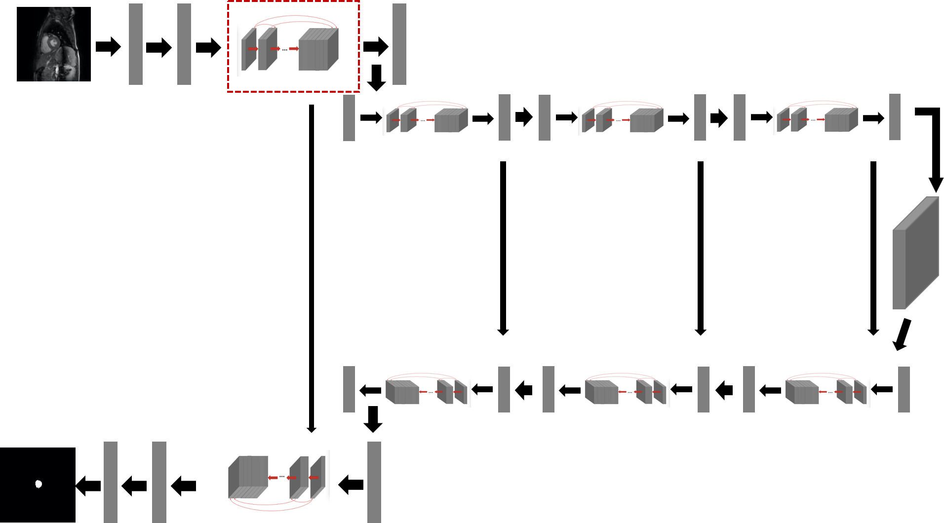
Network architecture: Figure 1 shows the network architecture of DenseV-Net that contains eight basic dense building blocks. The basic network architecture of DenseV-Net takes V-Net as reference [5],and the network can be easily divided into two parts. The first part is to use down convolutional layer to extract features of images and reduce the resolution of images with using different strides, so that the features can be compressed to be more fit for LV segmentation. The second part is to use up convolutional layers to decompress the signal and reconstruct the image until the output restored to original size. The down convolutional and up convolutional layers are all set appropriate parameters of padding. This network uses smaller memory footprint during training without switch mapping through convolutional and up-convolutional network. And it forwards the features extracted from compression part to the decompression part, avoiding loss of the features during compression path. Especially, ELUs is used to as active function.
Residual function: Residual function [4] is applied in basic dense building blocks (red block, Figure 1). It is utilized to alleviate the vanishing-gradient problem and strengthen feature propagation. This function is designed to be concatenated six times iteratively. Residual function ensures that every input of the convolutional layer in dense building block concatenates all the previous output of convolutional layer, which has proved to be able to relieve the increasing training error caused by increasing number of layers. In addition, we choose exponential linear units(ELUs) to add non-linearity as described [6]. Dice similarity coefficient (DSC) is used to evaluate the segmentation accuracy [4]. Segmentation performance of the proposed method is compared with that of conventional V-Net, with manual segmentation as reference.
Results
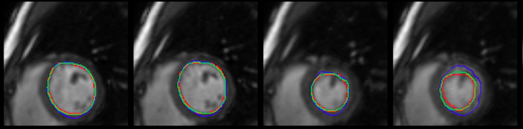
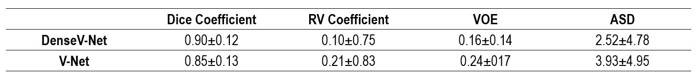
Compared to LV borders segmented with V-Net method (blue), the borders delineated with the proposed method (green) are found to be more comparable with those determined from manual tracing (red, Fig. 2). Linear regression analysis shows that the correlation between the proposed DenseV-Net and manual segmentation (R=0.92) is much stronger than that between V-Net and manual reference (R=0.75). Meanwhile, significantly higher mean DSC is observed for the proposed network (0.90±0.12) compared to that of V-Net (0.85±0.13, P<0.05, Table 1).Conclusion
The proposed DenseV-Net is a feasible method for automated LV segmentation with improved accuracy compared to the conventional V-Net method, providing a novel way for efficient heart function quantification from MR cardiac cines.Acknowledgements
This work was supported by the Guangdong Special Support Program (2017TQ04R395), the National Natural Science Foundation of China (81871441, 81571668, 81871348), the Natural Science Foundation of Guangdong Province in China (2017A030313743), the Shenzhen Overseas High-Level Talent Peacock Team of China (KQTD2016053117113327) and the Shenzhen Science and Technology Program (JCYJ20170307165550864, JCYJ20170413161350892).References
1. Yamamuro M, Tadamura E, Kanao S, Okayama S, Okamoto J, Urayama S, Kimura T, Komeda M, Kita T, Togashi K: Cardiac functional analysis by free-breath real-time cine CMR with a spatiotemporal filtering method, TSENSE: comparison with breath-hold cine CMR. J Cardiovasc Magn Reson 2006, 8(6):801-807.
2. Kaji S, Yang PC, B.kerr A, Tang WHW: Rapid evaluation of LV volume and mass without BH using real-time interactive CMR system. journal of the American College of Cardiology 2001(38(2):527-533).
3. Pham DL, Xu C, Prince JL: Current Methods In Image Mmedical Segmentation.
4. He K, Zhang X, Ren S, Sun J: Deep Residual Learning for Image Recognition. 2015.
5. Milletari F, Navab N, Ahmadi S-A: V-Net: Fully Convolutional Neural Networks Volumetric Medical Image Segmentation. 2016.
6. Clevert D-A, Hochreiter TUS: FAST AND ACCURATE DEEP NETWORK LEARNING BY EXPONENTIAL LINEAR UNITS (ELUS). 2016.
Figures