2222
USPIO-enhanced highly-resolved pulmonary MR angiography in mice with a 3D UTE sequence.Colleen R Cardiet1, Aurélien J Trotier1, Emeline J Ribot1, and Sylvain Miraux1
1CNRS - Univ. Bordeaux, CRMSB UMR 5536, BORDEAUX Cedex, France
Synopsis
Pulmonary imaging has always been challenging due to many constraints : parenchyma heterogeneity, respiratory and cardiac motion. Until now, pulmonary arteries imaging is commonly done with micro CT scanner, but this imaging method is not convenient for longitudinal evaluations. In this study, we present a magnetic resonance angiography (MRA) technique of the lungs with high spatial resolution combining an UTE 3D SG sequence and the injection of iron nanoparticles. To prevent motion artifact, images acquired during respiration were neglected.
INTRODUCTION
Magnetic resonance imaging (MRI) of pulmonary arteries has a significant role in the understanding of pulmonary and cardiac diseases. However, because of vessel size and respiratory motion, pulmonary angiography in small rodents is still challenging and requires high spatial and temporal resolutions. The reference method is micro CT scanner. However, this method is not suitable for longitudinal evaluation of pathologies. Pulmonary MR angiography (MRA) in living mice would therefore provide an important surrogate for cardiovascular research. This study aims at reconstructing MR angiography of mouse lungs with high spatial resolution, due to the use of a self-gated UTE 3D sequence with a pseudo-random encoding [1] combined with the injection of iron nanoparticles.METHODS
MRI experiments were performed on a 7T Bruker system (Germany), using a 4-phased array coil in a volume configuration (20mm diameter). C57BL/6 mice were anesthetized with isoflurane. Before mice positioning in the magnet, 100µmol Fe/kg of ultrasmall super paramagnetic iron oxide (USPIO, Ferumoxytol) was injected through the tail vein. A 3D self-gated UTE sequence was used : TR/TE=3.5/0.081 ms, Flip angle=15°, 5 data points sampled for self-gated signal (SG), 600 000 projections (30000 x 20 repetitions) distributing according to the pseudo Golden angle method, TA=35min. A field of view of 16x16x16 mm was used and a matrix size of 128x128x128 mm resulting in an isotropic resolution of 125 µm. From the SG data, the respiratory signal was extracted and 30 images were reconstructed along the respiratory cycle. Data during the respiratory movement were either not used to prevent motion artifact, and to improve spatial resolution (cases « 15 images » and « 1 image » in Figure 1), or used to show the impact of the respiratory movement on angiography images (case « 30 images » in Figure 1).RESULTS
When all the images acquire during the respiratory cycle are used, the signal increases but the spatial resolution decreases due to motion. Consequently, a compromise has to be made between the quantity of data from the respiratory peak used for reconstruction and the signal to noise ratio. Figure 2 shows the images obtained with three different mount of images, as described previously (30, 15 or 1 images). Image A illustrates that including the data acquired during the respiratory peak generates blurriness due to motion, compared to the one reconstructed with only the 15 images centered during the stable phase (B). In that latter case, a highly-resolved MR angiography was obtained, enabling the detection of small pulmonary vessels. Finally, using only one image of the respiratory cycle resulted in noise. Therefore, pulmonary angiographies were reconstructed using the 15 images centered during the stable phase. The image quality of lung parenchyma in our study is consistent (see figure 3), in which small blood vessels and their branches can be detected (red arrows), as well as bronchi (blue arrows).DISCUSSION
The present work shows the efficiency of using a 3D UTE SG sequence coupled with iron nanoparticles injection, to reconstruct highly-resolved pulmonary angiography of mice. By only taking data acquired during the stable phase of the respiratory cycle, image quality is improved. In 35 min, high quality angiography of the lungs, comparable to micro CT scanner, can be reconstructed, allowing small structure detection like vessels and bronchi.CONCLUSION
Here, we demonstrated the feasibility of providing highly-resolved free-breathing pulmonary angiography, with an isotropic spatial resolution of 125 µm in mice.Acknowledgements
No acknowledgement found.References
- Trotier AJ, Castets CR, Lefrançois W, Ribot EJ, Franconi JM, Thiaudière E, Miraux S. USPIO-enhanced 3D-cine self-gated cardiac MRI based on a stack-of-stars golden angle short echo time sequence : Application on mice with acute myocardial infarction.J Magn Reson Imaging. 2016 Aug. 44(2):355-65. doi: 10.1002/jmri.25150
Figures
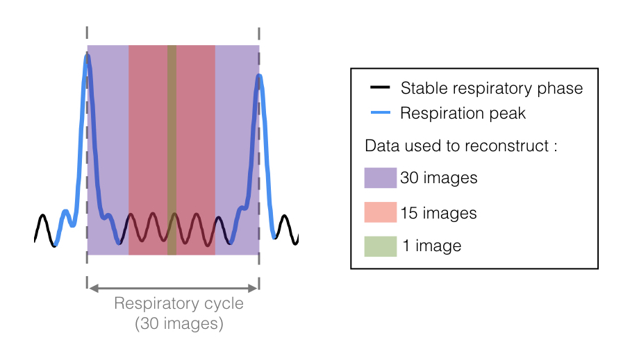
Self-gated signal of a free breathing mouse. Delimitations of images used for different reconstructions are shown in the scheme, to evaluate the impact of motion on the angiogram.
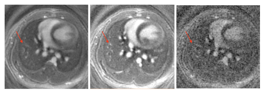
Data reconstruction with various numbers of cine images. Image A is reconstructed using all the data from the respiratory cycle, so 30 images. In B, we used 15 images, centered in the stable phase of the respiratory cycle. Finally, only the 15th image of the cycle was used in image C, explaining the low SNR. In B, the red arrow points out pulmonary vessels, that are not easily detectable in A and C.
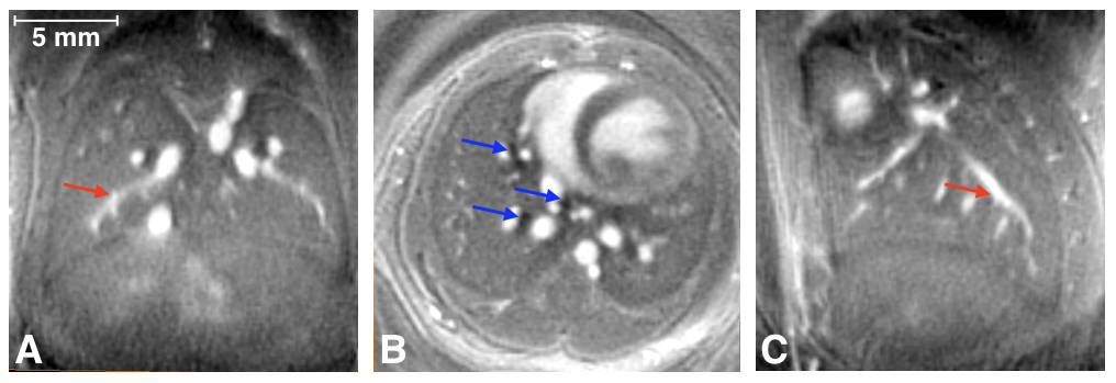
High-resolved free breathing pulmonary angiography. Oblique coronal (A), axial (B) and oblique sagittal (C) views of an angiography using 15 images by respiratory cycle. These images enable artifact-free and high-resolution depiction of the parenchymal lung structures, including small bronchi (black areas, shown by the blue arrows in B) and entire blood vessels (shown in A and B with the red arrows).