2220
Successfully Overcoming the Challenge: Cardiac Cine of Zebrafish in MRI.1MRI & µ-CT Service Group, Max-Planck-Institute for Heart and Lung Research, Bad Nauheim, Germany, 2Kerckhoff-Klinik, Kerckhoff-Klinik, Bad Nauheim, Germany, 3Bruker BioSpin MRI GmbH, Ettlingen, Germany, 4Department III Developmental Genetics, Max-Planck-Institute for Heart and Lung Research, Bad Nauheim, Germany
Synopsis
Zebrafish has been a widely used model to study cardiovascular development and diseases. Among the multiple advantages, zebrafish has the remarkable ability to regenerate lost or damaged organs including the heart. In vivo visualization of the cardiovascular system is particularly easy during early stages of development due to zebrafish transparency, which is lost in the adult stages. Therefore, it is of great interest to refine and adapt non-invasive imaging methods that allow a functional assessment of the zebrafish adult heart. The application of high-field functional MRI of the beating zebrafish heart will be presented and discussed.
Introduction
The zebrafish is a powerful model organism to study the development and regeneration of the cardiovascular system [1]. One of the main advantages of this animal model is the possibility to live track cellular and tissue dynamics during development using optical tools [2]. However, non-invasive live imaging of adult specimens is particularly challenging [3,4,5]. The objective of this study was to adapt the measurement parameters of cardiovascular magnetic resonance imaging (CMR) for an in vivo, non-invasive assessment of the cardiac function of the adult zebrafish heart.Methods and Materials
Measurements were performed on a
7T (Pharmascan, cryogenic 4 element array coil, receive-only,
and a room temperature transmit-only volume coil (72mm) small
animal MRI system. MRI
self-gating sequences [6] were used to monitor cardiac rate, opercular motion and sudden movements of the fish, for retrospective reconstruction of the cines.
Before scanning the zebrafish were anesthetized in a buffered 0.016% MS-222
solution (Sigma) until immobilization. For imaging zebrafish were placed in
supine position into the scanner. An overall view of an ex vivo zebrafish (Fig. 1) illustrates the heart chambers in a
sagittal view.Results
Successful cine reconstructions in various geometric orientations of the zebrafish heart were performed retrospectively by the evaluation of the selfgating signals. Monitoring the physiology of the zebrafish during data acquisition made it possible to identify the heart beat as well as sudden movements of the anesthetized fish. These occasional body movements could be discarded by the retrospective reconstruction. The navigators of the self-gated sequences were positioned such that they covered the ventricle, the bulbus arteriosus, the atrium and a combination of them. Spatially localized navigators could be used to derive the signal of each compartment individually. We were able to identify considerable variability in heart rate among individuals, but even more challenging for the reconstruction - an intrinsic variation in the same individual during the experiment.
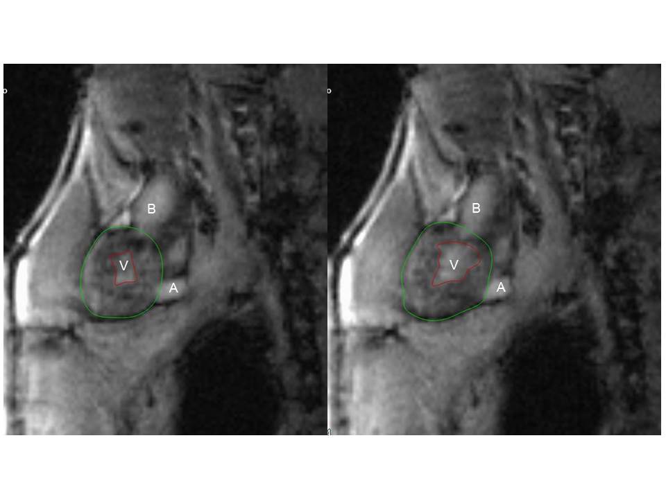
Fig. 2 shows the successful cardiac cine reconstruction of a two chamber view (ventricle and bulbus arteriosus) of a zebrafish whose heart rate switched several times between 70 and 130 bpm during measurement. Nevertheless the retrospective reconstruction provided excellent cines. In the three-chamber view (ventricle, atrium and bulbus arteriosus (Fig 3), one can depict the lumen of the ventricle.
Discussion
We have successfully acquired non-invasive functional in vivo imaging of the beating heart in adult zebrafish. Major limitations rise not only from the small anatomy demanding high-resolution, but also from the strong variation of the heart rate in some individuals, making conventional triggering impossible. The retrospective gating technique seems to be the only solution to overcome all these difficulties including triggering, sudden body movements and individual differences. Overall, we present a unique methodology to identify phenotypes affecting the morphology and, importantly, the function of the zebrafish adult heart. By the use of CMR we may now provide new insights on potential therapeutic approaches using zebrafish as a cardiovascular model.Acknowledgements
No acknowledgement found.References
[1] Stainier (2001). “Zebrafish genetics and vertebrate heart formation” Nat Rev Genet 2: 39-48.
[2] Mickoleit, M., et al. (2014). "High-resolution reconstruction of the beating zebrafish heart." Nat Methods 11(9): 919-922.
[3] Koth, J., et al. (2017). "High-Resolution Magnetic Resonance Imaging of the Regenerating Adult Zebrafish Heart." Sci Rep 7(1): 2917.
[4] Charles, J. F., et al. (2017). "Utility of quantitative micro-computed tomographic analysis in zebrafish to define gene function during skeletogenesis." Bone 101: 162-171.
[5] Merrifield, G. D., et al. (2017). "Rapid and recoverable in vivo magnetic resonance imaging of the adult zebrafish at 7T." Magn Reson Imaging 37: 9-15.
[6] Bovens SM et al. (2011). “Evaluation of infarcted murine heart function: comparison of prospectively triggered with self-gated MRI.” NMR Biomed 24: 307–315.
Figures
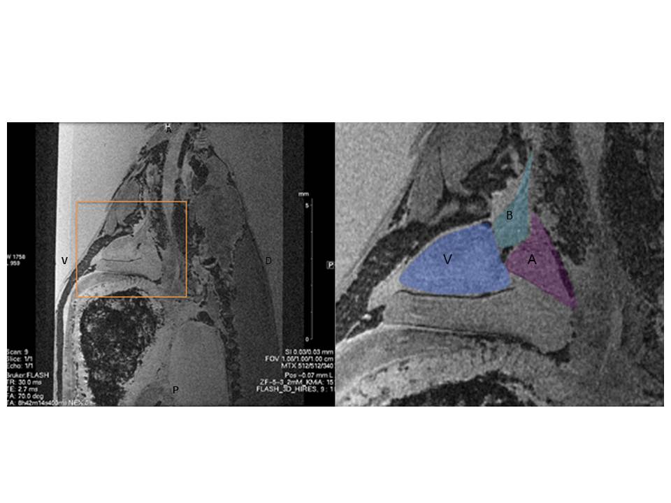
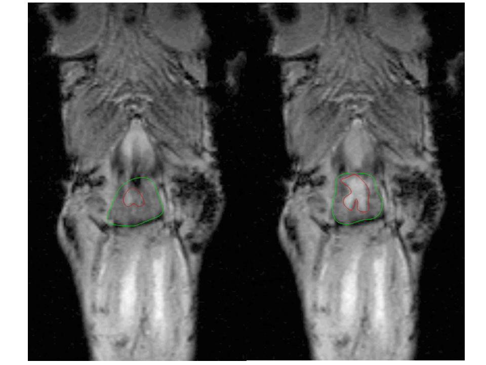
Fig. 2: Endsystolic (left) and enddiastolic (right) coronal views of bulbus arteriosus and ventricle of in vivo zebrafish heart. Intragate Flash (TE/TR = 4.2/5.2; flip angle = 10°; OS = 150; resolution = 59/59 µm)