2213
Changes of left ventricular myocardial strain in acute STEMI patients with microvascular obstruction: A CMR feature tracking study1Shengjing Hospital of China Medical University, Shen Yang, China, 2Shengjing Hospital of China Medical Univercity, Shen Yang, China
Synopsis
Changes of left ventricular myocardial strain in acute ST-elevation myocardial infarction patients before left ventricular remodeling is obvious, while the relation to microvascular obstruction still need to explore.
Objective
To characterize the alteration in quantification of global left ventricular (LV) strain by cardiovascular magnetic resonance(CMR) feature tracking after acute ST-elevation myocardial infarction(STEMI) before LV remodeling, and to explore the relation to microvascular obstruction (MVO).Methods
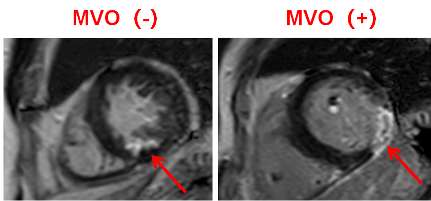
The prospective study enrolled 32 patients with STEMI successfully treated with primary PCI who underwent CMR after 3-5days (baseline) and 4-6 months (follow-up). Cine imaging and late gadolinium enhancement(LGE) was performed at 3.0 T MR. Longitudinal global strains(GLS),circumferential global strains (GCS),global radial strains(GRS) and cardiac volume were measured by CVI 42 software based on cine sequences. MVO was assessed based on LGE sequence. According to the existence of MVO or not, all patients were divided into MVO(+) group and MVO(-) group. Mean values of GLS, GCS and GRS were compared between the two groups of patients using an independent-samples t-test. Means of baseline and follow-up measurements were compared using a paired t-test.Results
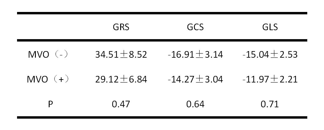
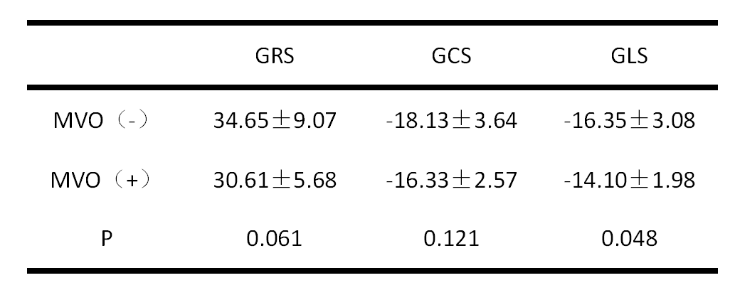
In this study, none of the patients had LV remodeling, according to the end-systolic volume improvement no more than 15%. Seventeen patients were classified as MVO(-) and 15 patients as MVO(+)[Age:(57.82±10.53)year vs.(55.07±10.42)year, P=0.774;BMI:(24.38±2.85)kg/m² vs.(27.06±3.13)kg/m²,P=0.698]. At baseline, there were no significant differences of GRS, GCS and GLS between the two groups (P=0.47,0.64 and 0.71). At follow-up, the GRS and GCS between MVO(+) and MVO(-) still make no significant differences (P=0.06, 0.12), while the GLS mean values of MVO(+) group significantly lower than that of MVO(-) group [(-14.10±0.51)%vs. (-16.35±0.75)%, P=0.048]. From baseline to 4-6 follow-up, the GRS, GCS and GLS of the two groups were all increased {MVO(-): [(34.51±8.52)% vs. (34.65±9.07)% , P =0.007 ], [(-16.91±3.14)% vs.( -18.13±3.64)% , P=0.038 ], [(-15.04±2.53)%vs. (-16.35 ±3.08)%,P=0.010];MVO(+):[(29.12±6.84)%vs. (30.61±5.68)% ,P=0.101], [(-14.27±3.04)%vs.( -16.33±0.66,)%, P=0.014 )], [(-11.97±2.21)%vs.(-14.10 ±1.98)% , P=0.657 ]}.Conclusions
For acute STEMI patients without LV remodeling in 4-6 months after PCI, the GRS, GCS and GLS of LV were all increased irrespective of MVO existed or not, and the changes of GLS in group with MVO was more obvious. Which may be the evidence of LV early remodeling.Acknowledgements
No acknowledgement found.References
1、Prediction of infarct size and adverse cardiac outcomes by tissue tracking-cardiac magnetic resonance imaging in ST-segment elevation myocardial infarction
2、Left ventricular long axis strain: a new prognosticator in non-ischemic dilated cardiomyopathy?
3、Prediction of functional recovery by cardiac magnetic resonance feature tracking imaging in first time ST-elevation myocardial infarction. Comparison to infarct size and transmurality by late
4、Longitudinal Strain Is a Marker of Microvascular Obstruction and Infarct Size in Patients with Acute ST-Segment Elevation Myocardial Infarction
Figures