2207
Diagnostic value of 3D T1-SPACE technique for basilar artery stenosis: Comparison with DSAzhenjia wang1, wei yu1, and zhaoyang fan2
1Department of Radiology, Beijing Anzhen Hospital, Capital Medical University, beijing, China, 2Biomedical Imaging Research Institute, Department of Biomedical Sciences, Cedars-Sinai Medical Center, Los Angeles, CA, United States
Synopsis
This is the first attempt to evaluate the accuracy
and application value of Three-Dimensional Variable-Flip-Angle Turbo Spin-Echo
(3D T1-SPACE) for the diagnosis of basilar atherosclerotic disease.
Introduction
To evaluate the accuracy and application value of Three-Dimensional Variable-Flip-Angle Turbo Spin-Echo [1-2](3D T1-SPACE) for the diagnosis of basilar atherosclerotic disease.Methods
54 patients with clinical symptoms suspected of having basilar artery disease were retrospectively evaluated with 3D T1-SPACE and DSA images. Basilar artery stenosis obtained from 3D T1-SPACE and DSA were independently determined. Spearman rank-order correlation coefficient was used to assess the correlation. Bland-Altman plots and intraclass correlation coefficient (ICC) were used to assess the agreement. The consistency rate, sensitivity, specificity, positive predictive value, negative predictive value, positive likelihood ratio and negative likelihood ratio were used to assess the diagnostic ability of 3D T1-SPACE technique.Results
A total of 54 patients (41 men, 13 women) received 3D T1-SPACE scanning and DSA successfully. 2 patients were excluded from the study due to unqualified image quality. There were excellent correlation and agreement between 3D T1-SPACE and DSA images in measuring luminal stenosis (r= 0.979, p<0.001; ICC, 0.99; 95% confidence interval [CI]: 0.98, 0.99). Bland-Altman plots showed that the two modalities were in good consistency in evaluating the degree of stenosis. The consistency rate, sensitivity, specificity, positive predictive value, negative predictive value, positive likelihood ratio and negative likelihood ratio of 3D T1-SPACE in diagnosing severe basilar artery stenosis were 92.3%,85.7%,96.4%,95.5%,90.0%,24.5 and 0.13, respectively.Discussion
Preliminary results from 54 patients demonstrated excellent image quality and high diagnostic value by 3D T1-SPACE technique. High-resolution 3D coronal acquisition allowed large coverage and flexible viewing which can provide much lesions informations. Some luminal blood signal inhomogeneity and T1 errors were likely due to inflow effects, especially at the vessel bifurcation.Conclusion
3D T1-SPACE technique is a noninvasive and accurate method in diagnosing basilar artery atherosclerotic disease.Acknowledgements
No acknowledgement found.References
[1] Fan Z, Zhang Z, Chung YC, et al. Carotid arterial wall MRI at 3T using 3D variable-flip-angle turbo spin-echo (TSE) with flow-sensitive dephasing (FSD). J Magn Reson Imaging. 2010. 31(3): 645-54.
[2] Fan Z, Zuehlsdorff S, Liu X, Li D. Prospective self-gating for swallowing motion: a feasibility study in carotid artery wall MRI using three-dimensional variable-flip-angle turbo spin-echo. Magn Reson Med. 2012. 67(2): 490-8.
Figures
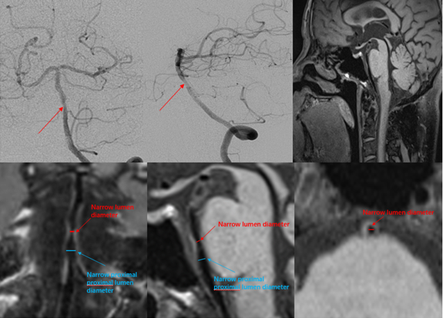
DSA and 3D T1-SPACE