2196
PolyGate: Benefits of a spatially resolved navigator for self-gating cardiac imaging in multiple rodents.Willy Gsell1,2, Cesar Molinos3, Carlos Correcher3, Michael Heidenreich4, Joren Vandengaer1, Wouter Oosterlinck5, Uwe Himmelreich1, Christophe M Deroose6, and Arno Nauerth7
1Biomedical MRI group, Department of Imaging and Pathology, KU Leuven, Leuven, Belgium, 2MoSAIC facility, Department of Imaging and Pathology, KU Leuven, Leuven, Belgium, 3Nuclear Molecular Imaging, Bruker BioSpin Preclinical Imaging Division, Bruker BioSpin, Valencia, Spain, 4Nuclear Molecular Imaging, Bruker BioSpin Preclinical Imaging Division, Bruker BioSpin, Ettlingen, Germany, 5Cardiac Surgery, department of cardiovascular Sciences, KU Leuven, Leuven, Belgium, 6Nuclear Medicine and Molecular Imaging, Department of Imaging and Pathology, KU Leuven, Leuven, Belgium, 7Nuclear Molecular Imaging, Bruker BioSpin Preclinical Imaging Division, Bruker Biospin, Ettlingen, Germany
Synopsis
We implemented a spatially resolved navigator based on the self-gated IntraGate-FLASH method. The new navigator offers many advantages such as the possibility to scan several animals at the same time, improve the quality of the motion correction and to distinguish between the different types of motion from a single acquisition. Moreover, we have been able to synchronize MRI with PET for simultaneous acquisition and to retrospectively reconstruct gated PET-images.
Introduction:
With depth of interaction (DOI) correction on a preclinical PET system, it is now possible to have homogenous resolution across the entire field of view1. Therefore, this opens the possibility of scanning multiple animals simultaneously to increase the throughput without compromising on the image data integrity. However, it is still challenging to perform cardiac imaging using ECG-gating or retrospective gating (IntraGate)2. We therefore implemented a new method, a spatially resolved navigator (PolyGate) enabling to extract the cardiac and respiratory motions of up to three separate animals at the same time and to retrospectively reconstruct cardiac gated PET data of multiple animals. But this new feature has much more to offer even for the acquisition of a single animal.Materials and Methods:
Here, we report on two cases for which the PolyGate method demonstrated added value compared to the already establish IntraGate-FLASH method for retrospectively gated MRI3. In all cases, PET-MRI data were acquired on a Bruker BioSpec 70/30 MRI system equipped with a SiPM based Albira PET insert (Bruker Biospin), using a quadrature volume coil (inner diameter of 86 mm). Case 1: Simultaneous acquisition of PET/MRI cardiac imaging in multiple animals. Three C57BL/6 mice of 25g were scanned simultaneously. They were all intravenously injected with 8MBq of 18F-FDG, one hour before the start of a 60 min static acquisition of PET data. Simultaneously, we acquired PolyGate-FLASH (TE/TR: 4/13.7 ms, 20 degrees flip angle, FOV: 70 x 30 mm, matrix 172×86 zero filled to 256 ×128, 3 slice packages of 6 slice of 1 mm thickness, 155 oversampling, flow compensated, total acquisition time: 54 min 48 s). The MRI sequence was modified to send a TTL signal to the PET DAQ electronics during the time of data collection at each TR loop. Then the spatially resolved IntraGate navigator information was used to derive the required retrospective data sorting scheme for each animal that represents the position within the cardiac cycle. The list-mode PET data were then rebinned according to the MRI based cardiac cycles to reconstruct 8 cardiac PET imaging frames using MLEM reconstruction with 0.25 mm isotropic resolution and 36 iterations. Case 2: Increase of the robustness of cardiac cine reconstruction compared to conventional IntraGate in a single animal. The same approach as detailed above was used with the following parameters (TE/TR: 4.2/14.5ms, 20 degrees flip angle, FOV: 30×30 mm, matrix 192×192, single slice package of 7 slices of 1 mm thickness, 250 oversampling, flow compensated, total acquisition time: 54 min 16 s). Animal was subject to permanent ligation of the left anterior descending artery and was scanned 2 weeks post ligation.Results:
Spatially resolved navigator (Fig.1) was successfully implemented, enabling us to acquire and reconstruct cardiac cine in three mice simultaneously (Fig.3). End systole and diastole were resolved in each animals in both the MRI and PET data. From the profile of the navigator signal, we could easily identified each animal and the fast acquisition combined with the increased quality of the navigator signal has even enabled us to monitor the respiration and cardiac rate in real time during the acquisition (Fig.2). In the second case, our self-gating approach has the benefit of improving the robustness of the cine reconstruction (98.3% of motion retrieval versus 82.8% using the full navigator signal) in infracted animals for which the motion may be altered (Fig.4). This resulted in sharper cine images.Conclusion and Discussion:
PolyGate and more specifically the introduction of spatially resolved navigators showed many benefits such as the possibility to scan multiple animals at the same time (increase of throughput) and increase of the robustness of the self-gated MRI sequence. This has not only implications for cardiac imaging but also benefits for other applications like liver and lung imaging in which motion correction is needed and can easily be implemented for clinical application. In some cases, arterial and venous phase can also be identified from the navigator signal providing a way to improve vascular imaging as well.Acknowledgements
No acknowledgement found.References
- A PET Detector Ring with Homogenous Spatial Resolution in the Presence of a Magnetic Field. Antonio J. González, Albert Aguilar, Andrea González-Montoro, Carlos Correcher, Pablo Conde, César Molinos, Konrad Lankes, Sven Junge, José M. Benlloch. IEEE 2015 Nuclear Science Conference Proceedings
- Retrospectively gated cardiac PET-MR imaging in rodents using MRI-based cardiac motion information. (#549). W. Gsell, A. Nauerth, C. Molinos, C. Correcher, A. J. Gonzalez, S. Sven, T. Greeb, R. Polo, B. Holvoet, C. M. Deroose, U. Himmelreich, M. Heidenreich. 13th annual meeting of the European Society for Molecular Imaging, ESMI. 20-23 March 2018.
- Evaluation of infarcted murine heart function: comparison of prospectively triggered with self-gated MRI. Bovens SM, te Boekhorst BC, den Ouden K, van de Kolk KW, Nauerth A, Nederhoff MG, Pasterkamp G, ten Hove M, van Echteld CJ. NMR Biomed. 2011 Apr;24(3):307-15.
Figures

Diagram of the PolyGate pulse sequence. The PolyGate
method has an added read gradient lobe in the navigator module. It results in
the generation of a signal profile in which 3 animals can easily be identified
(blue, pink and orange trace). The analysis of the signal changes over time of
the corresponding profile for each animal showed the respiratory (big drop of
signal amplitude) and cardiac (small changes) motion.

Spatially-resolved navigator signal for simultaneous acquisition
of cardiac cine in 3 mice. A: Display of the navigator signal where each
animal is assigned a color (animal1: blue, animal2:pink and animal3: orange).
In the top 3 tabs, the maximum intensity projection of the spectra for each
animal is displayed. B: Real time monitoring trace during the acquisition,
enabling the monitoring for respiration and cardiac rate.
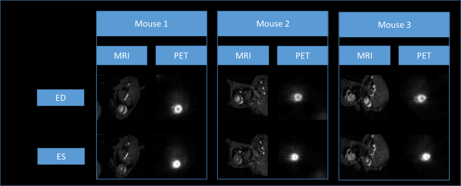
Case 1. Retrospective gated reconstruction of cardiac
cine for MRI and the use of the navigator signal to reconstruct PET data.
Cardiac frame at end diastole (ED) and end systole (ES) for animal 1, 2 and 3.
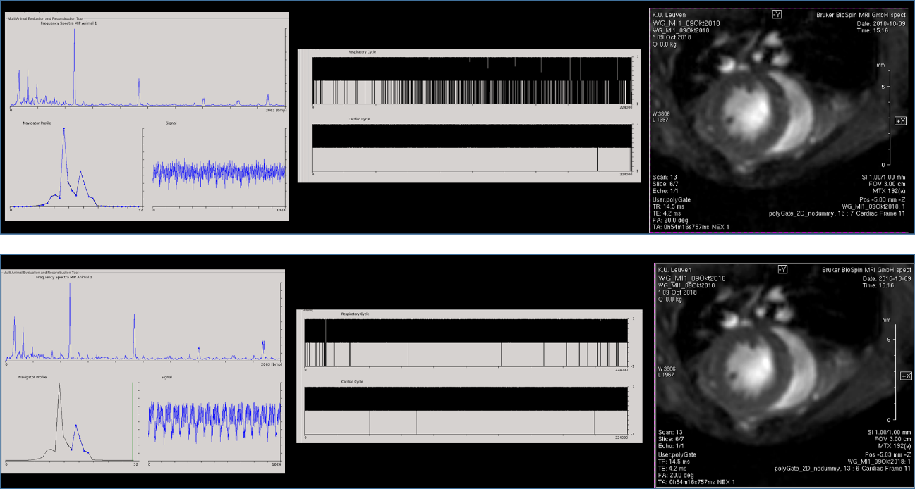
Case 2. Myocardial infarction in mice. The use of the
full navigator signal (top row) showed only 82.5% of motion retrieval mainly due
to missing respiration leading to a more blurry image compared to the selection
of the navigator profile where motion can be easily depicted (98.3% retrieval).