2192
3D Cartesian Free-running Cardiac and Respiratory Resolved Whole-heart MRI1School of Biomedical Engineering & Imaging Sciences, King's College London, London, United Kingdom, 2Siemens Healthineers, Erlangen, Germany
Synopsis
Free-breathing continuous acquisitions, so called free-running, enable 3D whole-heart coverage for motion-resolved functional cardiac MRI. In prior work approaches based on 3D radial imaging were proposed for this task. However, free-running 3D radial image reconstruction is computationally demanding. In this work, we propose a novel 3D Cartesian free-running bSSFP sequence. Data is acquired continuously with a variable-density Cartesian trajectory with spiral profile order and retrospectively binned based on respiratory self-navigation and cardiac ECG signal synchronization. A multi-bin-PROST reconstruction is proposed to exploit spatial, cardiac and respiratory temporal redundancies to reconstruct high-resolution images.
Introduction
3D radial1-4 free-breathing continuous acquisitions, so called free-running, have been recently proposed to reconstruct 3D whole-heart images at different cardiac and respiratory phases. These approaches retrospectively assign the data into different cardiac and respiratory phases using ECG or self-extracted cardiac and/or respiratory signals. Cardiac and respiratory resolved images are then reconstructed exploiting temporal redundancy in both cardiac and respiratory directions5. These approaches have shown promising image quality in reasonable scan times ~10-12min. Those methods1-7 however exhibit several drawbacks including long reconstruction times due to the non-Cartesian nature of the acquisition.
Here, we propose a 3D Cartesian free-running bSSFP acquisition which enables cardiac and respiratory resolved images of the whole-heart without contrast administration in a clinically feasible scan time and with adequate reconstruction times. A variable-density Cartesian sampling with spiral order (VD-CASPR)8,9 is extended to enable uniform retrospective sample-to-motion state binning. An embedded self-navigation signal captures the respiratory motion. A motion-resolved low-rank patch-based multi-bin-PROST reconstruction is proposed to exploit spatial, cardiac and respiratory temporal redundancies and to reconstruct high quality images.
Methods
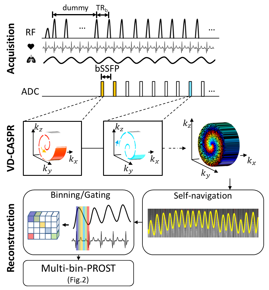
Acquisition: The proposed acquisition and reconstruction framework is depicted in Fig.1. In order to minimize eddy current artefacts, a VD-CASPR sampling with linear angle increment $$$\varphi$$$ and an outward-in trajectory between spirals continuously subsamples the Cartesian ky/kz plane by an acceleration factor L. Depending on L, matrix size and number of spiral rings, after S spirals a fully sampled k-space is reached, i.e. all high-frequency points are sampled at least once whilst the low-frequency range is oversampled. This property originates from the variable-density sampling and is desirable to enable low-frequency high-contrast sampling more frequently. The trajectory provides incoherent subsampling artifacts for the different motion-resolved images. Data is continuously acquired for a given desired acquisition time. The center line of k-space is periodically sampled serving as a 1D respiratory self-navigator. An α/2 preparation RF pulse and dummy pre-pulses bring the magnetization into bSSFP steady-state before data acquisition.
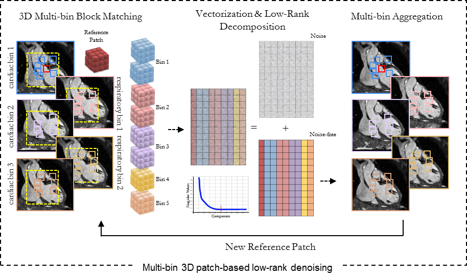
Reconstruction: A bandpass-filtered PCA extracts the respiratory-induced displacement of the heart from the 1D self-navigator. A retrospective dual gating bins the data into respiratory and cardiac states based on the respiratory self-navigation signal and ECG. Cardiac bins are adaptively adjusted per RR-interval to the current heart-rate to increase motion state consistency over all cardiac cycles. A parametrizable view-sharing amongst neighboring gates and a low-frequency view-sharing ensure good initialization for the subsequent reconstruction. The subsampled 6D k-space (3D spatial + 1 cardiac + 1 respiratory + Rx channels) is reconstructed extending PROST8 to a multi-bin-PROST for cardiac and respiratory motion-resolved reconstruction (Fig.2). PROST has been recently introduced to reconstruct subsampled MR images by exploiting the highly redundant information, on a local (similar patches within a neighborhood) and non-local (low rankness of all patches in the image) scale. Here the strong correlation shared between the multiple cardiac and respiratory images is also exploited by extending the search for similar patches to a spatial/cardiac-motion/respiratory-motion neighborhood. Multi-bin-PROST reconstruction iterates between an MR reconstruction optimization and an efficient patch-based denoising using an alternating direction method of multipliers.
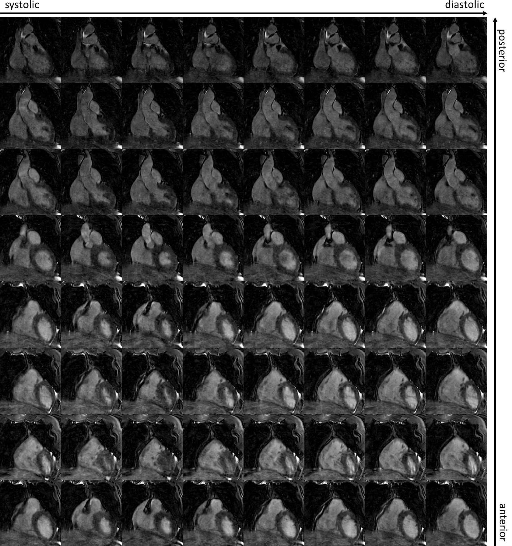
Data was acquired on a 1.5T MR (MAGNETOM Aera, Siemens Healthineers, Erlangen, Germany) in five healthy subjects in coronal orientation with 1.4mm3 isotropic resolution, FOV=320x320x260mm, TE/TR=1.6/3.2ms, flip angle=60°, spiral rings/segments=30, acceleration L=3, bandwidth=880Hz/px, 200 pre-dummy pulses, self-navigation period = 96ms, acquisition time = 7min52s. For the multi-bin-PROST a patch size of 5x5x5px, search window = 60px, patch offset = 4px with 20 simultaneously selected patches was used.
Results and Discussion
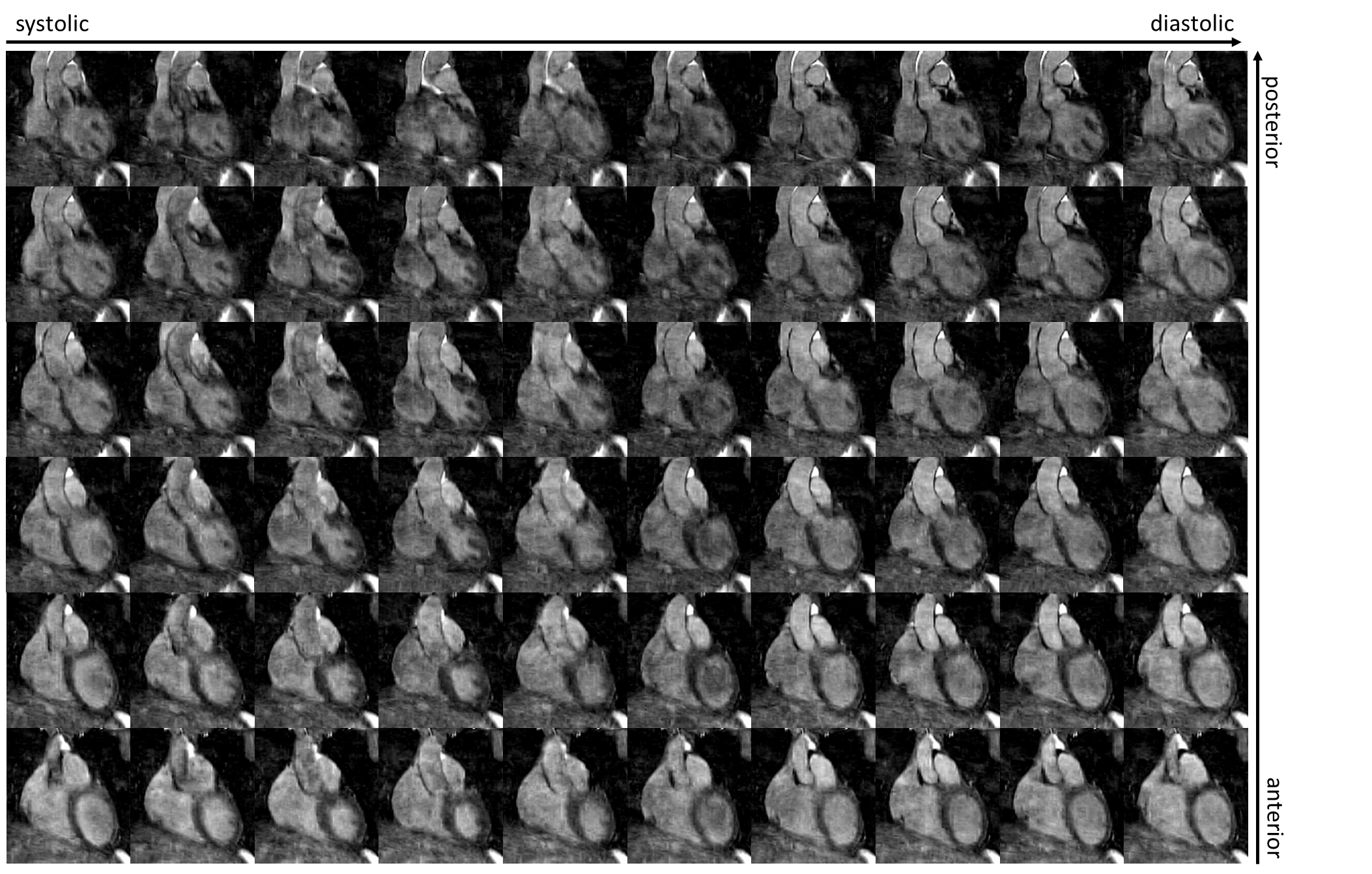
End-expiratory
cardiac motion-resolved images of two subjects obtained with the proposed
framework are shown in Figs.3-5. Images indicate a good spatial and temporal
resolution for whole-heart coverage in a reasonable scan time (~8min). Gating
can effectively reduce motion-induced blurring. Sufficient image contrast was
obtained in the heart for the continuous free-running acquisition. Linear angle
increment and spiral out-inward trajectory reduces eddy current-induced
artifacts in the image compared to a golden angle increment. Exploiting spatio-temporal
redundancy via multi-bin-PROST recovers high-resolution motion-resolved images.
Small moving structures such as the right coronary artery can be visualized
(Fig.3).Conclusion
The proposed acquisition provides 3D Cartesian cardiac and respiratory resolved images with high spatial and temporal resolution under free-breathing in ~8min scan time. Multi-bin-PROST reconstruction fully exploits the spatio-temporal redundancies in the acquired data to reconstruct high-resolution images in ~5min/state reconstruction time. Future studies will investigate further acceleration of the proposed acquisition and the application of a fast-interrupted steady-state (FISS)10 to enable intrinsic fat suppression.Acknowledgements
This work was supported by EPSRC (EP/P007619) and Wellcome EPSRC Centre for Medical Engineering (NS/A000049/1).References
1. Coppo S, Piccini D, Bonanno G, Chaptinel J, Vincenti G, Feliciano H, Van Heeswijk RB, Schwitter J, Stuber M. Free‐running 4D whole‐heart self‐navigated golden angle MRI: initial results. Magnetic resonance in medicine 2015;74(5):1306-1316.2. Pang J, Sharif B, Fan Z, Bi X, Arsanjani R, Berman DS, Li D. ECG and navigator‐free four‐dimensional whole‐heart coronary MRA for simultaneous visualization of cardiac anatomy and function. Magnetic resonance in medicine 2014;72(5):1208-1217.
3. Wu HH, Gurney PT, Hu BS, Nishimura DG, McConnell MV. Free‐breathing multiphase whole‐heart coronary MR angiography using image‐based navigators and three‐dimensional cones imaging. Magnetic resonance in medicine 2013;69(4):1083-1093.
4. Liu J, Nguyen TD, Zhu Y, Spincemaille P, Prince MR, Weinsaft JW, Saloner D, Wang Y. Self-gated free-breathing 3D coronary CINE imaging with simultaneous water and fat visualization. PloS one 2014;9(2):e89315.
5. Feng L, Coppo S, Piccini D, Yerly J, Lim RP, Masci PG, Stuber M, Sodickson DK, Otazo R. 5D whole-heart sparse MRI. Magnetic resonance in medicine 2018;79(2):826-838.
6. Han F, Rapacchi S, Khan S, Ayad I, Salusky I, Gabriel S, Plotnik A, Finn JP, Hu P. Four‐dimensional, multiphase, steady‐state imaging with contrast enhancement (MUSIC) in the heart: A feasibility study in children. Magnetic resonance in medicine 2015;74(4):1042-1049.
7. Usman M, Ruijsink B, Nazir M, Cruz G, Prieto C. Free breathing whole-heart 3D CINE MRI with self-gated Cartesian trajectory. Magnetic resonance imaging 2017;38:129-137.
8. Prieto C, Doneva M, Usman M, Henningsson M, Greil G, Schaeffter T, Botnar RM. Highly efficient respiratory motion compensated free-breathing coronary MRA using golden-step Cartesian acquisition. Journal of magnetic resonance imaging : JMRI 2015;41(3):738-746.
9. Bustin A, Ginami G, Cruz G, Correia T, Ismail TF, Rashid I, Neji R, Botnar RM, Prieto C. Five-minute whole-heart coronary MRA with sub-millimeter isotropic resolution, 100% respiratory scan efficiency, and 3D-PROST reconstruction. Magnetic Resonance in Medicine 2018.
10. Küstner T, Bustin A, Jaubert O, Neji R, Prieto C, Botnar R. 3D Cartesian Fast-interrupted Steady-state Sequence (FISS) with Intrinsic Fat Suppression. submitted to. Proceedings of the International Society for Magnetic Resonance in Medicine (ISMRM)2019. p 1511.
Figures