2182
One-stop scanning of cerebrovascular, carotid and cardiovascular MRI with a 48-channel coil system at 3 T1Lauterbur Imaging Research Center, Shenzhen Institutes of Advanced Technology, Chinese Academy of Sciences, Shenzhen, China, 2Shanghai United Imaging Healthcare, Shanghai, China, 3Department of Radiology and Biomedical Imaging, University of California San Francisco, San Francisco, CA, United States, 4UCSF/UC Berkeley Joint Graduate Group in Bioengineering, San Francisco, CA, United States
Synopsis
One-stop
magnetic resonance (MR) vessel wall imaging that includes cardiovascular
magnetic resonance (CMR), intracranial and cervical carotid arteries imaging is
of considerable
interest for clinical diagnosis of vascular diseases, such as ischemic stroke.
Due to the small cross-sectional size of the vessel wall and susceptibility
effects, one-stop MR vessel wall imaging remains challenging. In this work, intracranial
and cervical carotid arteries, and CMR imaging was performed simultaneously by
using a dedicated 48-channel coil system. Moreover, intracranial and carotid
arterial wall images with an isotropic spatial resolution of 0.63 mm can be
acquired by using the head and carotid coil system within 7.7 minutes.
Introduction
In ischemic stroke, atherosclerotic plaque is the main cause, which is found to be prevalent in cardiovascular, carotid and cerebrovascular vessels1. However, for plaque detection, most studies were focused on the intracranial and cervical carotid arteries2, and some studies mainly on craniocervical artery dissection3. Simultaneous intracranial and extracranial arterial wall imaging would greatly enhance the capability and convenience of clinical diagnosis for plaque, which can be achieved by magnetic resonance imaging (MRI) with vessel wall imaging or black blood imaging technology. In this work, a one-stop scanning of cerebrovascular, carotid and cardiovascular MRI was implemented by using a proposed 48-channel coil system at a 3 T MRI system (uMR 790, Shanghai United Imaging Healthcare, Shanghai, China). The intracranial and carotid arterial wall image quality acquired by the head and carotid coil system was compared with that acquired by a commercially available head and neck joint coil array (17-channel head coil and 7-channel neck coil).Methods
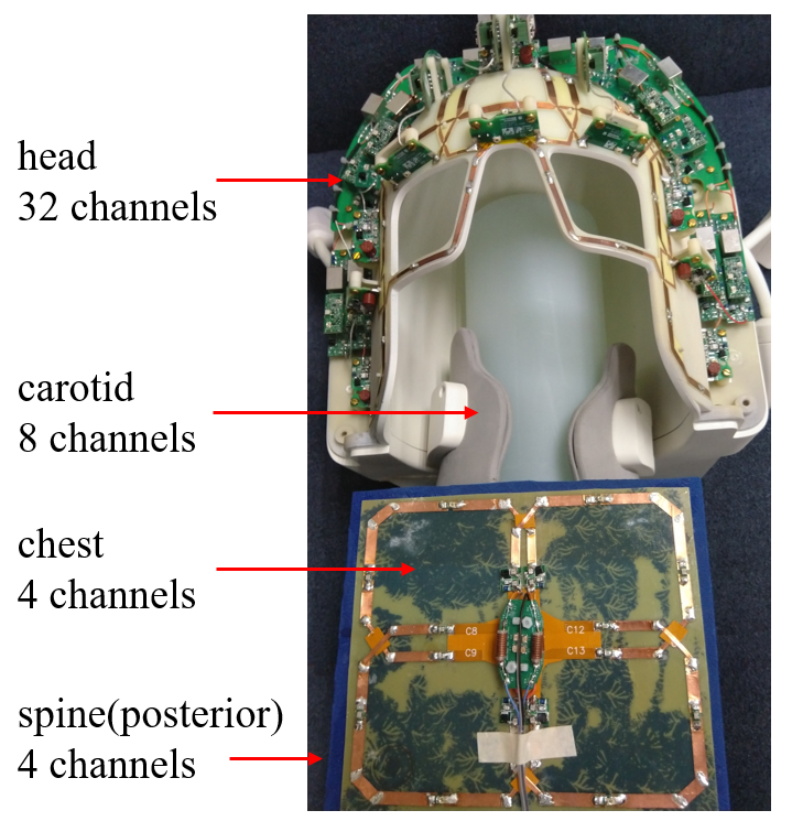
To accurately identify the intracranial and carotid arterial plaques, the spatial resolution must reach 0.5 mm isotropy for that the thickness of intracranial and carotid arterial wall4 is less than 1 mm, while a 1 mm spatial resolution can satisfy the requirement of the thoracic aortic plaques diagnosis as the normal thickness of thoracic aortic vessel wall5 is close to 2 mm. Based on these spatial resolution requirements and the percentage of plaque distribution (46.6% in the intracranial artery, 27.9% in the carotid artery, and 25.5% in the heart and thoracic aorta), a 48-channel coil system consisting of a 32-channel head coil, an 8-channel carotid coil, a 4-channel chest coil and a 4-channel spine coil, was built for one-stop MR vessel wall imaging.
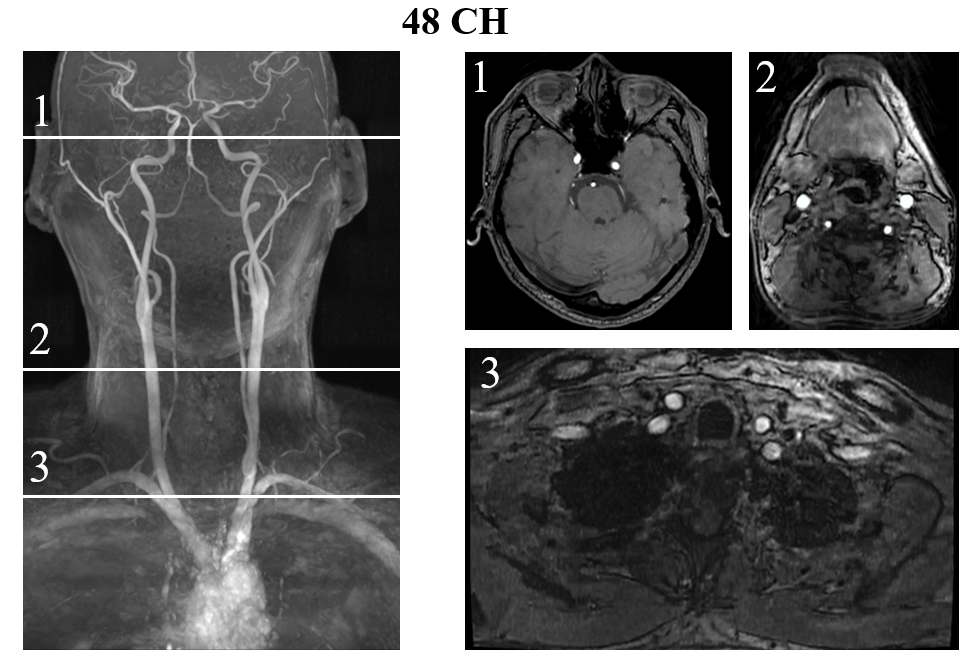
The human studies were IRB approved, and informed consents were obtained from all subjects. For MR angiography (MRA), 3D time-of-flight (TOF) imaging was applied to illustration of imaging coverage with the following parameters: turbo field echo, TR/TE= 20 ms/4 ms, flip angle= 16o, bandwidth= 250 Hz/pixel, slice thickness= 5 mm, slice number= 620, FOV= 220 mm (A–P) × 310 mm (S–I), and voxel size=1.07 × 0.86 × 1 mm3, total time=10.87 min. Here, the 48-channel coil system was applied.
For MR vessel wall imaging, a modified 3D MUST-MATRIX (MUST: Motion induced attenutation by Unbalanced STeady-state free precession preparation; MATRIX: Modulated flip Angle Technique in Refocused Imaging with eXtended echo train) sequence was applied with parameters: TR/TE=850 ms/14.7 ms, echo train length=40, FOV=212 mm (S-I) x 192 mm (L-R), slice thickness=0.6 mm, slices=240, matrix size=336 x 304, Bandwidth=600 Hz/Px, accelerate factor=3.5. Fat suppression was applied in the sequence. Here, the scan of cerebrovascular, carotid and cardiovascular vessel wall need to be done in two parts: one scan of cerebrovascular and carotid vessel wall, and another scan of cardiovascular vessel wall with the need to control breathing artifacts and heartbeat artifacts. The scan of cerebrovascular and carotid vessel wall took 7.7 min with an isotropic spatial resolution of 0.63 mm x 0.63 mm x 0.6 mm. In this process, both the head and carotid coil system and the head and neck joint coil array were applied. For the scan of cardiovascular vessel wall, the 4-channel chest coil and the 4-channel spine coil can be used.
Results
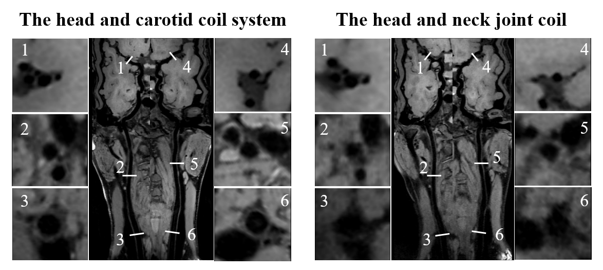
The 3D TOF MRA image obtained from the 48-channel coil system is shown in Fig. 2, from which it can be seen that cardiovascular, carotid and cerebrovascular vessel can be acquired in one-stop with a large FOV. The MR vessel wall images acquired by the head and carotid coil system and the head and neck joint coil array are shown in Fig. 3. As indicated by the arrows, the vessel wall irregularities along the middle cerebral artery are much sharper in the image acquired with the proposed the head and carotid coil system (see inserts). For cerebrovascular imaging, respiratory navigation and ECG-gating need to be applied. This work is under optimization and will be shown in the future.Conclusion
In conclusion, one-stop scanning of cerebrovascular, carotid and cardiovascular MRI can be achieved by using the proposed 48-channel coil system at 3T. The MR vessel wall images can also be clearly visualized, demonstrating its potential for clinical diagnosis.Acknowledgements
This work was supported in part by NSFC under Grant No. 61571433, 61801466, 81627901, 81527901; Guangdong Province grants 2014A030312006, and 2014B030301013; Youth Innovation Promotion Association of CAS No. 2017415; city grants JCYJ20170413161314734; NIH U01EB023829, and a Pengcheng Scholar Award.References
1. Z. Zhou, R. Li, X. Zhao, L. He, X. Wang, J. Wang, N. Balu, and C. Yuan, “Evaluation of 3D multi-contrast joint intra- and extracranial vessel wall cardiovascular magnetic resonance,” Journal of Cardiovascular Magnetic Resonance, vol. 17, no. 41, pp. 1-11, May. 2015.
2. X. Hu, Y. Li, L. Zhang, X. Zhang, X. Liu, and Y. C. Chung. “A 32-channel coil system for MR vessel wall imaging of intracranial and extracranial arteries at 3T,” Magnetic resonance imaging, vol.36, pp. 86-92, Feb. 2017.
3. X. Zhao, H. R. Underhill, Q. Zhao, J. Cai, F. Li, M. Oikawa, L. Dong, H. Ota, T. S. Hatsukami, B. Chu, and C. Yuan. “Discriminating carotid atherosclerotic lesion severity by luminal stenosis and plaque burden,” Stroke; a journal of cerebral circulation, vol.42, no.2, pp. 347-353, Feb. 2011.
4. Y. Qiao, D. A. Steinman, Q. Qin, M. Etesami, M. Schär, B. C. Astor, and B. A. Wasserman. “Intracranial arterial wall imaging using three-dimensional high isotropic resolution black blood MRI at 3.0 Tesla,” Journal of Magnetic Resonance Imaging, vol.34, no.1, pp. 22-30, Jul. 2011.
5. B. Mensel, A. Quadrat, T. Schneider, J. Kühn, M. Dörr, H. Völzke, W. Lieb, K. Hegenscheid, and R. Lorbeer. “MRI-based Determination of Reference Values of Thoracic Aortic Wall Thickness in a General Population,” European Journal of Radiology, vol. 4, no. 9, pp. 2038-2044, Sep. 2014.
Figures