2173
Role of Cardiac magnetic resonance imaging in evaluation of left ventricular function and myocardial stress difference in patients with hypertrophic cardiomyopathy1Ningxia Medical University, Yinchuan, China, 2Ningxia Medical University General Hospital, Yinchuan, China
Synopsis
Cardiac magnetic resonance multi-parameter imaging was used to evaluate left ventricular function and myocardial stress in patients with hypertrophic cardiomyopathy. A series of multiparametric magnetic resonance imaging was performed on 34 patients with asymmetrical hypertrophic cardiomyopathy diagnosed by MRI, using CVI software. Cardiac function was detected, and 322 hypertrophic myocardial segments and 222 non-hypertrophic segments were characterized for post-tracking. According to the American Heart Association 17-segment rule, the strain parameters and left ventricle of 16 segments except the apex were measured. The overall strain parameters preliminarily concluded that the left ventricular global stress of patients with hypertrophic cardiomyopathy was lower than that of normal people; the peak radial strain, peak long axis strain, peak circumferential strain and peak diameter of hypertrophic myocardial segments in patients with hypertrophic cardiomyopathy The strain rate and peak circumferential strain rate were significantly lower than the non-hypertrophic myocardial segments.
Objective
To evaluate left ventricular function and myocardial stress in patients with hypertrophic cardiomyopathy using cardiac magnetic resonance imaging.
Methods
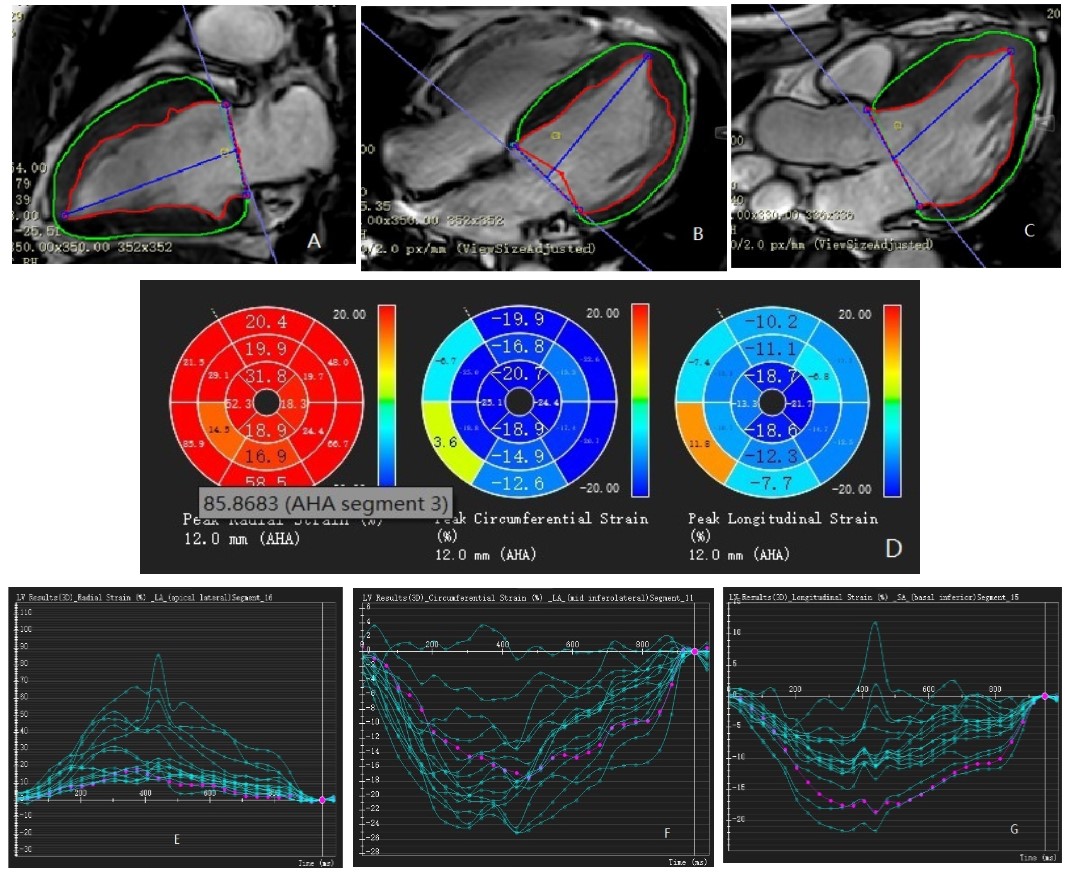
:Phlipis 3.0T Ingenia magnetic resonance instrument, ECG, respiratory gating technique and free steady-state sequence were used to obtain the standard cardiac short-axis two-chamber heart, long-axis two-chamber heart, four-chamber heart and inflow and outflow channel level, vertical 8-10 layers were continuously scanned from the apex to the short axis of the heart at room septum and as parallel as possible to the mitral valve. Scanning parameters: TR=2.9, TE=1.43, flip angle (FA) is 45°, FOV is 350*350, layer thickness is 8mm, pitch is 0, and 30 frames of movie images are acquired for each cardiac cycle. Cardiac function was detected using CVI software, thereby obtaining left ventricular function parameters, including LV end-diastolic volume (LVEDV), left ventricular end-systolic volume (LVESV), left ventricular effusion ejection fraction (LVEF%); and 34 patients with left ventricular global radial strain (RS), long axis strain (LS) and circumferential strain (CS), and The 322 hypertrophic myocardium segments and 222 non-hypertrophic segments were subjected to a signature tracking post-processing, and the peak radial strain (pRS), peak longitudinal strain (pLS), peak circumferential strain (pCS), and radial strain rate, longitudinal strain rate, Peak circumferential strain rate of 16 segments except the apex was measured according to the American Heart Association 17-segment rule.
Results
There were 37 patients with HCM, 3 of whom were excluded due to lack of image quality or left ventricular short axis. The standard HCM patients were 34 patients, including 28 males and 6 females. The average age was ( 45.3 ± 13.27) and (56.3 ± 15.92) years old. The range of male left ventricular cardiac function parameters LVEDV, LVESV and LVEF were (151.47, 48.74; 107.92) ml, (64.40, 10.08; 31.08) ml and (85.03, 50.71)%; female left ventricular cardiac function parameters (156.01, 60.15; 73.20) ml, (93.51, 14.11; 22.52) ml and (74.37, 60.06; 68.67)%. The left ventricular global radial strain (RS), longitudinal strain (LS) and circumferential strain (CS) of patients with hypertrophic cardiomyopathy were significantly lower than the left ventricular global radial strain (RS) and length of normal healthy Chinese adults. Longitudinal strain (LS) and circumferential strain (CS) [(34.02±16.57)% vs (79.0±19.94)%, p=0.000, (-19.98±5.46)% vs (-22.4±2.9)%, p=0.015, (-9.10 ± 5.05)% vs (-24.3 ± 3.1)%, p = 0.000]. The peak radial strain (pRS), peak longitudinal strain (pLS) and peak circumferential strain (pCS) of hypertrophic myocardial segments in patients with hypertrophic cardiomyopathy were significantly lower than those in non-hypertrophic myocardial segments [(15.31±9.45)vs(46.48±20.29)%, p=0.000;(-9.96±7.78) vs (-8.38±5.49)%, p=0.000; (-21.75±7.29) vs (-13.89±7.11)%, p =0.006]. The peak radial strain rate and peak circumferential strain rate of hypertrophic myocardial segments in patients with hypertrophic cardiomyopathy were lower than those in non-hypertrophic myocardium [(3.30±1.82) s-1 vs (1.16±0.95) s-1; (-1.58±0.76) s-1 vs (-1.13±0.86) s-1, p<0.05]. The peak long-axis strain rate of hypertrophic myocardial segments in patients with hypertrophic cardiomyopathy was not significantly different from that of non-hypertrophic myocardial segments [(3.30±1.82)s-1 vs(1.16±0.95)s-1, p=0.66] .Conclusions
The global left ventricular stress in patients with hypertrophic cardiomyopathy is reduced. For patients with hypertrophic cardiomyopathy with left ventricular function >50%, CMR-Tissue tracking can detect changes in myocardial strains in hypertrophic myocardium, and changes in hypertrophic myocardial segmental stress can be found earlier than left ventricular function parameters. Abnormal ventricular contractile function.Acknowledgements
-References
1. Normal Values of Myocardial Deformation Assessed by Cardiovascular Magnetic Resonance Feature Tracking in a Healthy Chinese Population: A Multicenter Study Junping Peng1,2,3, Xiaodan Zhao4, Lei Zhao2, Zhanming Fan2, Zheng Wang2, Hui Chen2, Shuang Leng4, John Allen5, Ru-San Tan4,5, Angela S. Koh4,5, Xiaohai Ma2*, Mingwu Lou3* and Liang Zhong4,5* Physiology doi: 10.3389/fphys.2018.01181
2. Augustine, D., Lewandowski, A. J., Lazdam, M., Rai, A., Francis, J., Myerson, S.,et al. (2013). Global and regional left ventricular myocardial deformation measures by magnetic resonance feature tracking in healthy volunteers: comparison with tagging and relevance of gender. J. Cardiovasc. Magn. Reson.15:8. doi: 10.1186/1532-429X-15-8
3.The comparison of short-term prognostic value of T1 mapping with feature tracking by cardiovascular magnetic resonance in patients with severe dilated cardiomyopathy Rui Chen1,2 · Jingjing Wang1,2 · Zhicheng Du3 · Yu‑Hsiang Juan4 · Carmen Wing‑Sze Chan5 · Hongwen Fei6 · Jiajun Xie7 · Wanjia Wu2 · Yulei Zhu1,2 · Liwen Li6 · Jinxiu Meng6 · Shulin Wu6 · Changhong Liang1,2 · Zhuliang Yu1,8 · Hui Liu1,2,6 The International Journal of Cardiovascular Imaging 2018.8
4.Kowallick, J. T., Morton, G., Lamata, P., Jogiya, R., Kutty, S., Lotz, J., et al. (2016). Inter-study reproducibility of left ventricular torsion and torsion rate quantification using MR myocardial feature tracking. J. Magn. Reson. Imaging 43, 128–137. doi: 10.1002/jmri.24979
Figures