2170
CMR coupled to optical flow and virtual fields methods for the quantification of myocardial mechanical properties in cancer survivors.1Polytechnique Montreal, Montreal, QC, Canada
Synopsis
Subtle changes in strains and mechanical proprieties of the myocardial tissue were assessed in childhood acute lymphoblastic leukemia survivors using the optical flow and virtual field methods on CMR images. Significant changes between risk groups were observed on the strain in the 2 chambers view only. However, significant changes between risk groups were observed on the stresses, shear and Young’s moduli in all views. The dynamic elastic and shear moduli obtained along the cardiac cycle might be indicators of doxorubicin induced cardiotoxicity.
Introduction
Doxorubicin-based chemotherapy is effective and widely used to treat acute lymphoblastic leukemia (ALL). However, its effectiveness is hampered by cardiotoxic effects depending primarily on the cumulative dose administered (1). The optical flow is an iconic method studying the apparent motion of objects by considering that the movement between images is determined based on the pixel gray level. The virtual field method is an inversion method dedicated to the problem of identification of mechanical properties from kinematic field measurements. This approach has been shown to be insensitive to borderline boundary uncertainties and robust, and has been successfully applied to different materials with non-linear behavior such as elastomers and biological tissues (2). The objective of this study was to assess the subtle changes in the mechanical properties of the left ventricle in ALL survivors exposed to doxorubicin associated or not to a cardioprotective agent.Methods
Nine ALL survivors were prospectively included and divided into 3 groups according to their prognostic risk: standard risk (SR, n=3), high risk (HR, n=3) and high risk group who received dexrazoxane, a cardioprotective agent (HRdex, n=3). Healthy subjects (n=3) from an in-house study were also included. They all underwent a CMR acquisition including an ECG-gated cine TruFISP sequence at 3T (Siemens SkyraTM) using a 18-channel phased array body matrix coil. The myocardium extraction was done using an interactive implementation of Bezier curves (1). The optical flow was implemented considering the conservation of data, which assumes that the intensity of the pixel remains constant over time, and the spatial coherence, which is based on the consistency of the movement between two pixels of the same neighborhood. For the two-chambers (2CH), four-chambers (4CH), and short-axis (AX) views, the Von Mises and shear strain were averaged over the systole, early diastole, and late diastole. We proposed a virtual field method based on the principle of virtual works. The only contribution that we considered was the virtual work done by the external forces. The relation between stress and strain in the elastic isotropic regime allowed the estimation of the Young's modulus and shear modulus for the systole, early diastole, and late diastole. One-way analysis of variance was performed between the four groups (HR, HRdex, SR, HV).Results
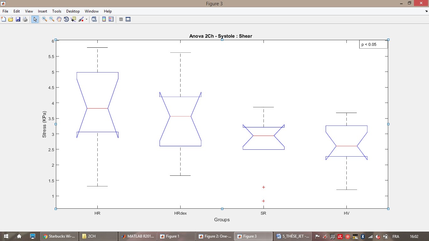
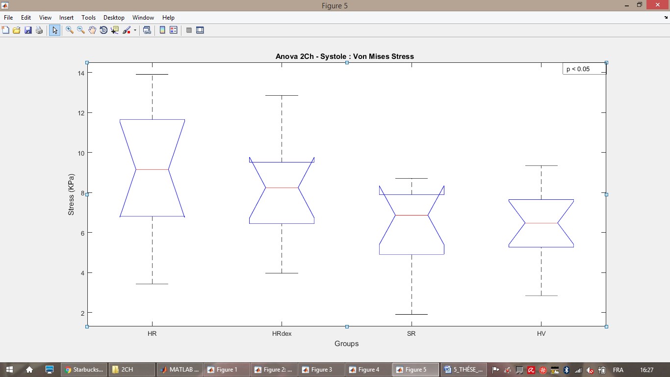
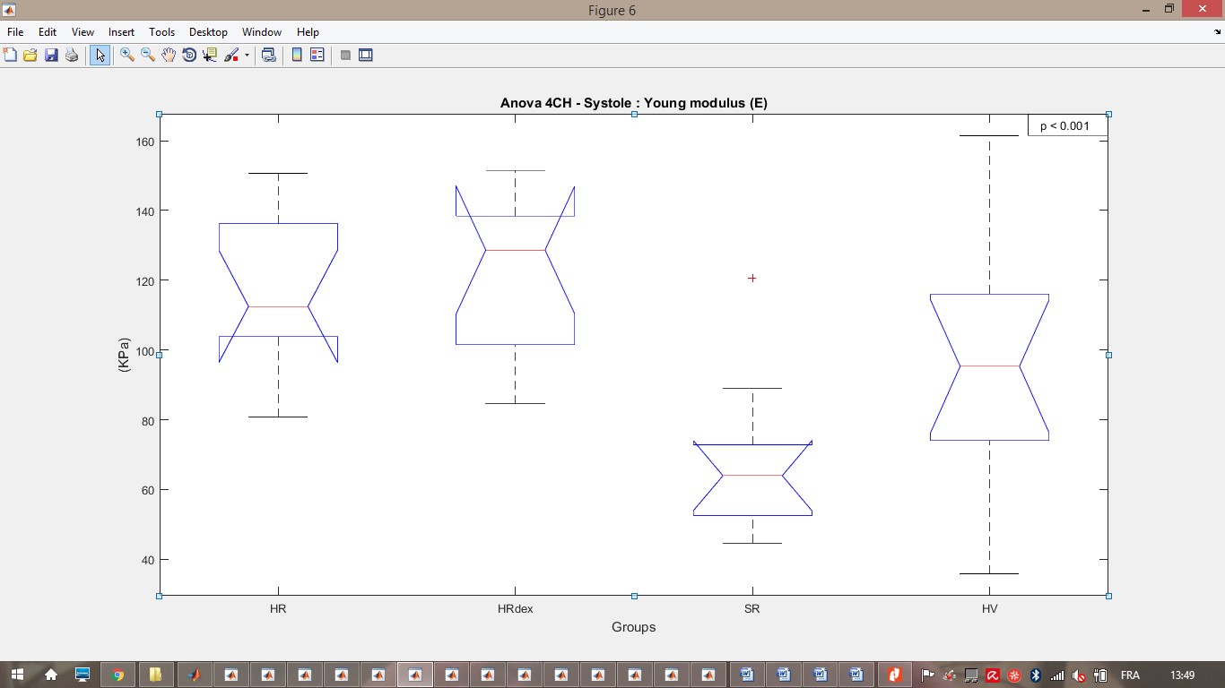
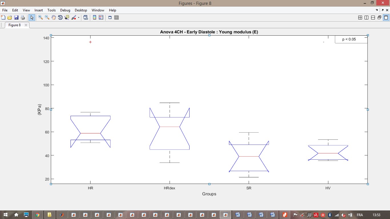
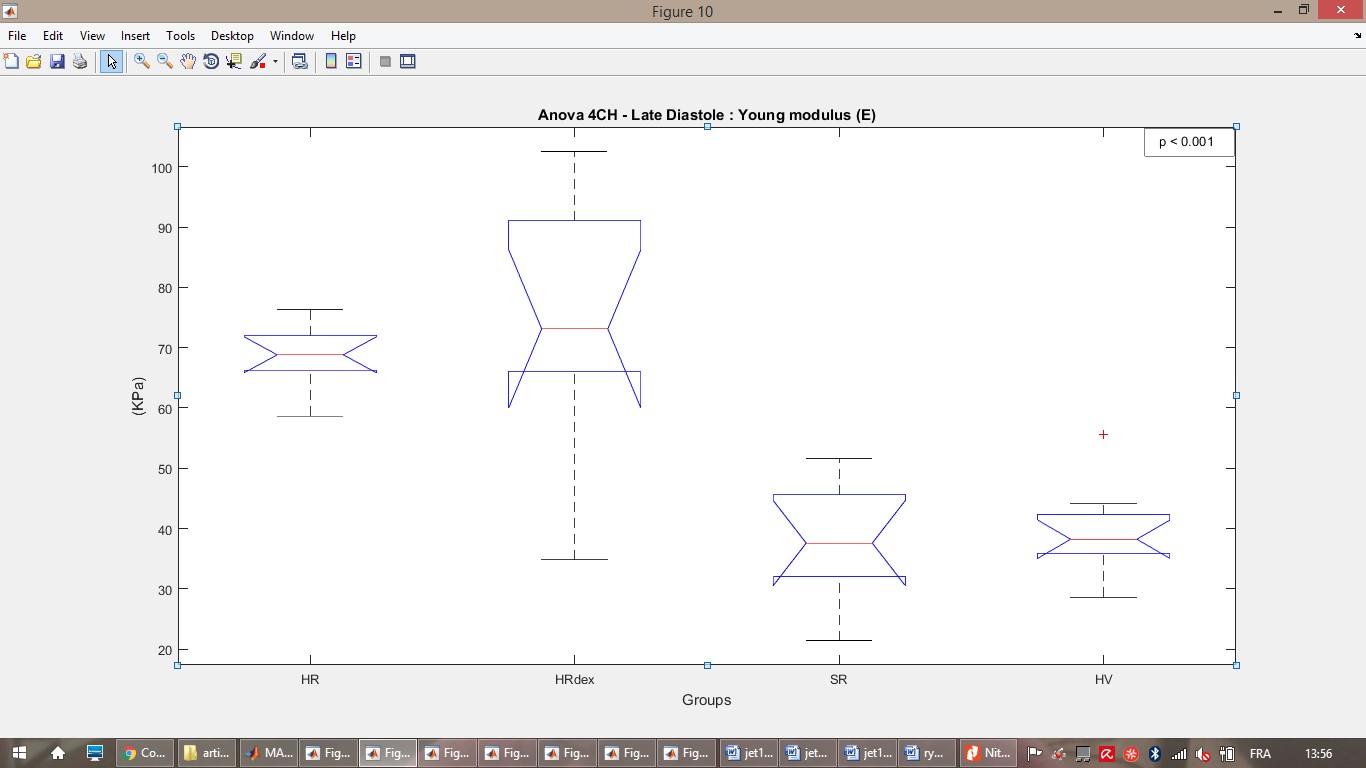
In 2CH, Von Mises strain were reduced in HRdex as compared to HR for late diastole only while shear strain were reduced in HRdex as compared to HR, SR and HV for both systole, early diastole and late diastole (p< 0.01). No significant differences were found in 4CH or AX views (p > 0.05). In 2CH, shear and Von Mises stresses were significantly reduced during systole in SR as compared to HR, HRdex and HV, and also reduced in HRdex as compared to HR (Figures 1 and 2, p<0.05). In 4CH, the Young’s modulus was significantly increased in HR and HRdex as compared to HV during all the cardiac cycle (Figures 3 to 5, p<0.05 ). In AX, shear stresses, Von Mises stresses and Young’s modulus were increased in HRdex as compared to HR, and decreased in HV as compared to SR and HRdex for both systole and late diastole (p<0.05).Discussion
Decreased shear strain and increased stiffness in HR and HRdex groups correspond to an increase of the amount of the muscle fibers in the myocardium and a decrease of the interstitial tissue (3). This is in agreement with fibrosis observed with doxorubicin induced cardiotoxicity. Groups of survivor whose modulus of elasticity is greater tends to deform less easily, in agreement with observed dilated cardiomyopathy immediately following high-dose anthracycline treatment (4,5).The optical flow method gives a field of displacements based only on pixel gray levels, without any interpolation. However, the method requires the same image resolution for all images. Assuming external contribution only and quantifying the virtual strain using the CMR strains simplified the application of the virtual field method. The dynamic elastic modulus obtained along the cardiac cycle included both passive and contractile behaviors. The low number of subjects included in each group limited this study to a feasibility study.Conclusion
Subtle changes in myocardium remodeling due to childhood cardiotoxicity can be accessed from the mechanical behavior analysis of the myocardial tissue based on cine-MRI images, the optical flow method and the virtual field method. Our approach allowed the quantification of the internal stresses and elastic modulus at each pixel of the cine-MRI image, reflecting the nature of myocardial tissue changes even in the same region of interest. The next modelling step will be to incorporate the contribution of the work done by the volumetric and acceleration forces.Acknowledgements
NSERC and Polytechnique Montreal for the financial support, researchers from the PETALE study for the opportunity to do this complementary analyses on the cancer survivors.References
1- Singal PK, Iliskovic N. Doxorubicin-induced cardiomyopathy. New England Journal of Medicine 1998; 339(13):900-905.
2- Bersi MR, Bellini C, Di Achille P, Humphrey JD, Genovese K, Avril S. Novel Methodology for Characterizing Regional Variations in the Material Properties of Murine Aortas. J Biomech Eng. 2016; 138(7).
3- Aissiou M1, Périé D, Cheriet F, Dahdah NS, Laverdière C, Curnier D. Imaging of early modification in cardiomyopathy: the doxorubicin-induced model. Int J Cardiovasc Imaging. 2013; 29(7):1459-76.
4- Lipshultz S, Sallan S. Cardiovascular abnormalities in long-term survivors of childhood malignancy. Journal of Clinical Oncology 1993;11(7):1199-1203.
5- Lipshultz SE, Lipsitz SR, Sallan SE, et al. Chronic progressive cardiac dysfunction years after doxorubicin therapy for childhood acute lymphoblastic leukemia. Journal of clinical oncology 2005;23(12):2629-2636.
Figures