2166
Multi-shot compressed sensing techniques accelerate cine sequence acquisition: an evaluation of diagnostic efficacy1Radiology, Peking Union Medical College Hospital, Beijing, China, 2Application, Siemens Shenzhen Magnetic Resonance Ltd., Shenzhen, China, 3Siemens Healthcare GmbH, Erlangen, Germany
Synopsis
An evaluation of diagnostic efficacy study about compressed sensing techniques accelerates cine sequence acquisition, which quantitatively and qualitatively compares 3 kinds of cine sequences in CMR.
Objective
Cardiac magnetic resonance cine images are conventionally acquired in breath-hold with a segmented balanced steady state free precession (bSSFP) sequence, which requires a relatively long acquisition time and high patient cooperation. Single-shot compressed sensing (ss CS) cine is a real-time sequence with reasonable spatial and temporal resolution that can be applied free-breathing[1]. However, the contrast between myocardium and surrounding soft tissue is relatively reduced and the epicardial delineation is not very accurate[2]. In this study, we used multiple shot compressed sensing cine technique to quickly acquire high-quality images and accurately assess cardiac function.Methods
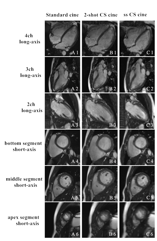
22 patients underwent cardiac MRI on a 3T scanner (MAGNETOM Skyra, Siemens Healthcare, Erlangen, Germany). For all patients, cine imaging was performed using three different methods: standard segmented cine sequence(8-shot), ss CS cine sequence, and prototype 2-shot compressed sensing (2-shot CS) cine sequence[3]. The key parameters of these sequences are shown in Table 1. We scan 4 chamber, 3 chamber and 2 chamber view for long axis, and 8-10 slices cover all left ventricle for short axis. Quantitative analysis of image quality(0-4 score system) and cardiac function were performed on cine images acquired from different sequences. All quantitative values were compared in a correlation study utilizing Spearman correlation analysis. Statistical significance was determined using Wilcoxon signed-rank test, and values p<0.05 were considered significant.Results
22 patients completed three types of cine sequences. The average scan time of standard cine sequence was (102.50±18.55)s, the average scan time of ss CS cine was (20.50±3.71)s, and the average scan time of 2-shot CS cine was (30.75±5.57)s. The standard cine sequence image score was (3.68±0.64), the ss CS cine sequence image score was (3.13±0.35), and the 2-shot cine sequence image score was (3.54±0.51), . In terms of quantitative study of cardiac function, the differences between the standard cine sequence and the ss CS cine sequence were no statistically significant except for image score and LV mass, as specific results shown in Table 2. There were no statistical differences in cardiac function parameters between the standard cine sequence and the 2-shot cine sequence. In terms of correlation study, there was a good correlation between standard cine and ss CS cine, and standard cine and 2-shot CS cine (P<0.01), as specific results shown in Table 3.
Conclusion
Compared with a single-shot cine sequence, a 2-shot compressed sensing cine sequence can acquire images closer in quality to the standard cine sequence but with a higher scan speed and with acceptable accuracy.Acknowledgements
No acknowledgement found.References
1. Vincenti G, Monney P, Chaptinel J, Rutz T, Coppo S, Zenge M, et al. Compressed sensing single-breath-hold CMR for fast quantification of LV function, volumes, and mass.[J]. Jacc Cardiovascular Imaging, 2014, 7(9):882-892.
2. Kido T, Nakamura M, Watanabe K, Schmidt M, Forman C. Compressed sensing real-time cine cardiovascular magnetic resonance: accurate assessment of left ventricular function in a single-breath-hold[J]. Journal of Cardiovascular Magnetic Resonance, 2016, 18(1):50.
3. Forman C, Kroeker R, Schmidt M., Accelerated 2D Cine MRI Featuring Compressed Sensing and ECG-Triggered Retro-Gating, ISMRM 2017, pp. 4669, 2017.
Figures
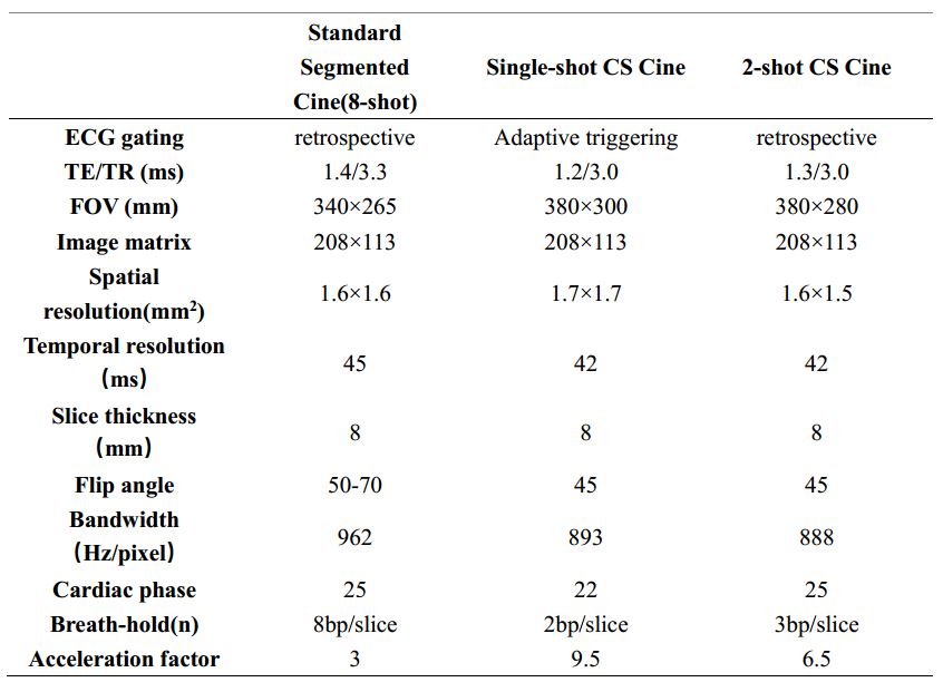
Table 1 Imaging parameters
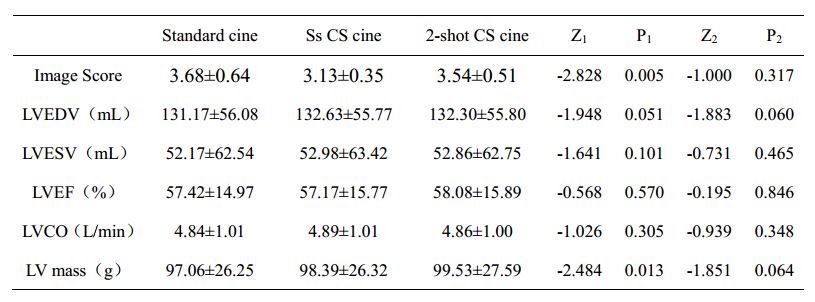
Table 2 Quantitative comparison of cardiac function results between sequences
* Note: Since the normality test results show that the pairwise difference is non-normally distributed, the Wilcoxon signed-rank sum test was used for statistical analysis. The Z values are based on the negative rank. Z1 and P1 are the pairing results between standard cine and ss CS cine; Z2 and P2 are the pairing results between standard cine and 2-shot CS cine.
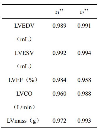
Table 3 Cardiac function parameter correlation study
*Note: Since the normality test results show a non-normal distribution, Spearman correlation analyses were used. r1 values are the correlation results between standard cine and ss CS cine; r2 are the correlation results between standard cine and 2-shot CS cine.