2159
Demonstration of Circumferential Heterogeneity in Displacement and Strain in the Abdominal Aortic Wall by Spiral Cine DENSE MRI1Department of Medicine, Emory University, Atlanta, GA, United States, 2Emory University, Atlanta, GA, United States, 3Atlanta VA Medical Center, Atlanta, GA, United States, 4Siemens, Atlanta, GA, United States
Synopsis
Herein, we describe a method for implementation of cine DENSE imaging in the abdominal aortic wall. The method uses in house software to post-process and track displacement of intermural pixels and applies a quadrilateral based solution to measure strain. This new method permits the measurement of circumferential heterogeneity of both displacement and strain.
Introduction
Vascular kinematics are predictors of cardiovascular mortality (1) but the ability to measure relevant parameters using MRI is insufficient for clinical utilization. Current MRI techniques used to measure vascular displacement and strain are unable to resolve circumferential heterogeneity. (2) The ability to quantify regional kinematics in the aorta would constitute a significant contribution given that heterogeneity is critical in rupture risk stratification for abdominal aortic aneurysms (AAA) for which rupture location depends on local, patient-specific biomechanics (3). Displacement-encoding with stimulated echoes (DENSE) is an MRI sequence capable of quantitatively measuring sub-pixel displacement and strain in the vessel wall (4). The objective of this study was to apply a time-resolved, 2D, spiral cine DENSE imaging sequence to measure displacement of the infrarenal aorta wall to assess baseline heterogeneities in radial and circumferential displacement, and to use this displacement data to quantify circumferential strain. We hypothesized that both displacement and circumferential strain would significantly differ around the circumference of the vessel.Methods
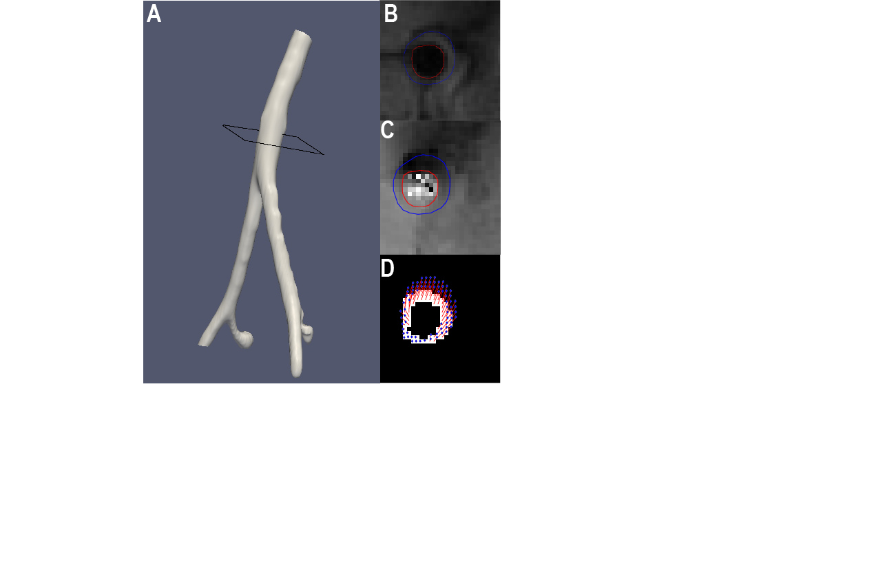
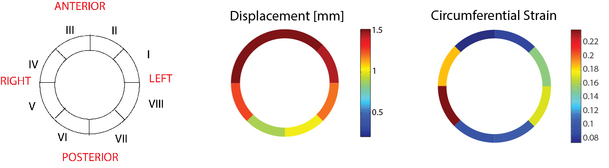
Seventeen subjects (n=9 women, n=8 men) with no cardiovascular disease were recruited for the IRB-approved study. For each subject, a prospectively ECG-triggered and free-breathing navigator-echo gated, spiral cine DENSE sequence was used to acquire a slice perpendicular to the aorta midway between the renal artery origin and the aortic bifurcation (Figure 1A). The sequence used segmented, spiral k-space sampling with displacement encoding value (ω) of 0.25 cycles/mm ,TE of 1.25 ms, TR of 16 ms, 8 spiral interleaves per image, 1 spiral interleave per heartbeat, slice thickness of 8 mm, and reconstructed pixel size of 1.8× 1.8mm (Figure 1B). Sequential acquisitions were performed to encode displacement in the two directions of the image plane (Figure 1C) and post processing was performed using a custom DENSE analysis program written in Matlab.. Using each of the phase image sets, displacement fields were created for the vessel wall (Figure 1D) and converted to polar coordinates. Green strain was calculated in the image coordinate system using an interpolation method based on quadrilateral elements(5) and converted to a polar coordinate system. For each volunteer, peak displacement and stain in each pixel was assigned to one of 8 normalized sectors (Figure 2A) to quantify and visualize regional heterogeneity while minimizing noise. Comparisons between sectors mean displacement and strain were made with a linear mixed model. An additional phase contrast cine image was acquired in the same location as the DENSE image and distensibility was calculated as $$$R^2/r^2-1$$$, where R is the radius at peak systole and r is the radius at end diastole. Distensibility was compared with homogenized DENSE-derived strain using Bland Altman analysis.Results
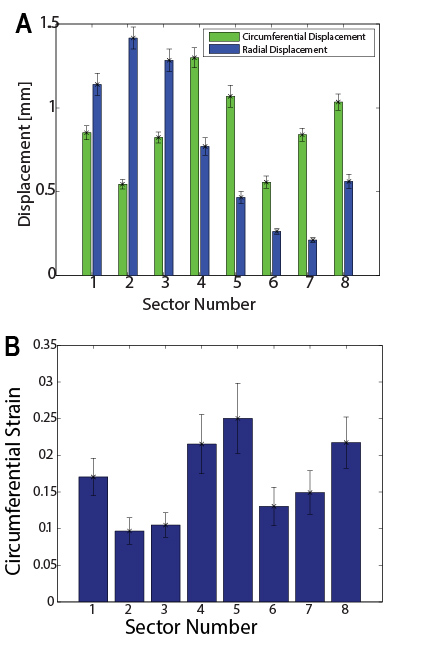
Average peak displacement was greater in the anterior vessel compared with the posterior half (1.5 ± 0.7mm vs 0.9±0.6 mm, p<0.05, Figure 2B). Sectors I-III (left anterolateral and anterior walls) had larger radial than circumferential displacement (p<0.01, Figure 3A), but sectors IV-VIII (right anterolateral through left posterolateral wall) demonstrated greater circumferential displacement (p<0.01). Peak circumferential strain (Figure 2C) in the anterior wall was lower than in each of the lateral walls (p<0.05 comparing anterior to left and p<0.05 comparing anterior to right, Figure 3B). The mean (i.e., homogenized) circumferential strain was calculated for all volunteers to be 0.14±0.05. Correlation between distensibility and homogenized strain was evaluated with a Pearson correlation coefficient of 0.76 and Bland Altman analysis demonstrated an average bias of 0.005±.0050.Discussion
This work demonstrates that cine DENSE imaging can be used to determine regional displacement and circumferential strain in the abdominal aorta. Application of this technique shows that while displacement is greatest in the anterior wall, circumferential strain is greatest in the lateral walls. These findings suggest posterior fixation of the aorta (low displacement and low strain) with displacement of the anterior wall in “bulk” (high displacement but low strain). Thus, the circumferential stretching of the aortic wall to is accomplished by stretch of the lateral walls (moderate displacement, high strain). The value of the proposed method’s ability to measure displacements with sub-pixel resolution is important when considering the potential for clinical application.Conclusion
Spiral cine DENSE imaging is able to resolve heterogeneities in aortic kinematics. We demonstrate that 2D cine DENSE imaging in the vessel wall is feasible in healthy volunteers and we report significant differences in the displacement and circumferential strain around the vessel circumference. The kinematics of the aorta are heterogeneous, even in young healthy adults which may have implications in understanding susceptibilities of distinct regions to the development and progression of pathologies of the infrarenal aorta, such as AAAs, in whichevolving biomechanical properties of the extracellular matrix likely play a significant role.Acknowledgements
The work in this abstract was supported by a pre-doctoral fellowship from the American Heart Association anda grant from Siemens Healthineers.References
1. Cavalcante J, Lima JA, Redheuil A, Al-Mallah MH. Aortic stiffness: current understanding and future directions. Journal of American College of Cardiologists 2011;5(57):1511-1522.
2. Suever J, Oshinski JN, Rojaas-Campos E, Huneycutt D, Cardarelli F, Stillman AE, Raggi P. Reproducibility of pulse wave velocity measurements with phase contrast magnetic resonance and applanation tonometry. Internation Journal of Cardiovascular Imaging 2011.
3. Martufi G, Christian Gasser T. Review: the role of biomechanical modeling in the rupture risk assessment for abdominal aortic aneurysms. Journal of Biomechanical Engineering 2013;135(2).
4. Aletras A, Ding S, Balaban RS, Wen H. DENSE: Displacement Encoding with Stimulated Echoes in Cardiac Functional MRI. Journal of Magnetic Resonance 1999;137:247-252.
5. JD H. Cardiovascular Solid Mechanics: Cells, Tissues, and Organs. New York, NY: Springer: 2002.
Figures