2147
Deep Learning for Radial SMS Myocardial Perfusion Reconstruction1Biomedical Engineering, University of Utah, Salt Lake City, UT, United States, 2UCAIR, University of Utah, Salt Lake City, UT, United States, 3Radiology and Imaging Sciences, University of Utah, Salt Lake City, UT, United States, 4Cardiology, University of Utah, Salt Lake City, UT, United States
Synopsis
Although dynamic contrast enhanced MRI has been successfully applied for characterizing coronary artery diseases, an acquisition scheme limited to 2-4 short axis slices restricts coverage of the left ventricle. Radial simultaneous multi-slice has been shown to improve DCE cardiac perfusion by providing complete coverage of the left ventricle but also requires an increase in reconstruction time. Here we propose using a modified Unet with a residual artifact learning framework to improve reconstruction time and image quality of spatio-temporal constrained reconstruction methods for radial SMS datasets. Results demonstrate promising improvements with a speed up in reconstruction by a factor of ~150.
Introduction
Dynamic contrast enhanced MRI has been used to better differentiate between different soft tissue structures of the heart and has allowed for increased detection and characterization of different coronary artery disease states. Dynamic contrast enhanced MRI of the heart is typically conducted through the acquisition of 2-4 short axis slices. However, this acquisition scheme provides incomplete coverage of the left ventricle of the heart and so provides limited information of coronary artery disease states such as ischemia in the myocardium. Radial simultaneous multi-slice (SMS) has been shown to improve DCE cardiac perfusion by providing complete coverage of the left ventricle [1]. However, the increase in data undersampling from radial SMS also increases the reconstruction time required. To improve image quality and reconstruction time, we used deep learning neural networks with a residual learning framework in order to learn the iterative pixel tracking spatio-temporal constrained reconstruction (PT-STCR) with total variation constraints [2-4]. Improved image quality and reconstruction times are obtained by learning the residual artifacts from the PT-STCR reconstructed images.Methods
Radial SMS data was obtained from 12 subjects who were scanned at rest and/or stress, free breathing, with or without ECG gating. Multiple sets of radial SMS data were acquired with each cardiac cycle. Each SMS set sampled three parallel slices that were either short axis slices, two chamber long axis slices, or four chamber long axis slices [5,6]. Radial SMS data were processed to suppress streaking [6] and principal component analysis (PCA) was used to compress the number of coils in the SMS data [6]. SMS data were then interpolated onto Cartesian space using extended GROG [7]. A preliminary STCR reconstruction with 30 iterations was conducted to create reference images for self gating and respiratory state binning. A novel motion robust pixel tracking framework was employed with STCR (PT-STCR) to create reconstructed SMS images [8]. All SMS data were acquired on Siemens 3T scanners. All radial data with golden ratio based spacing shared similar parameters as follows: TR = 2.7 ms, TE = 1.6 ms, FOV = 260 mm, ~1.8x1.8x8mm pixel size, 30 rays/frame, gadoteridol dose ~ 0.075 mmol/kg per injection and flip angle = 12 .
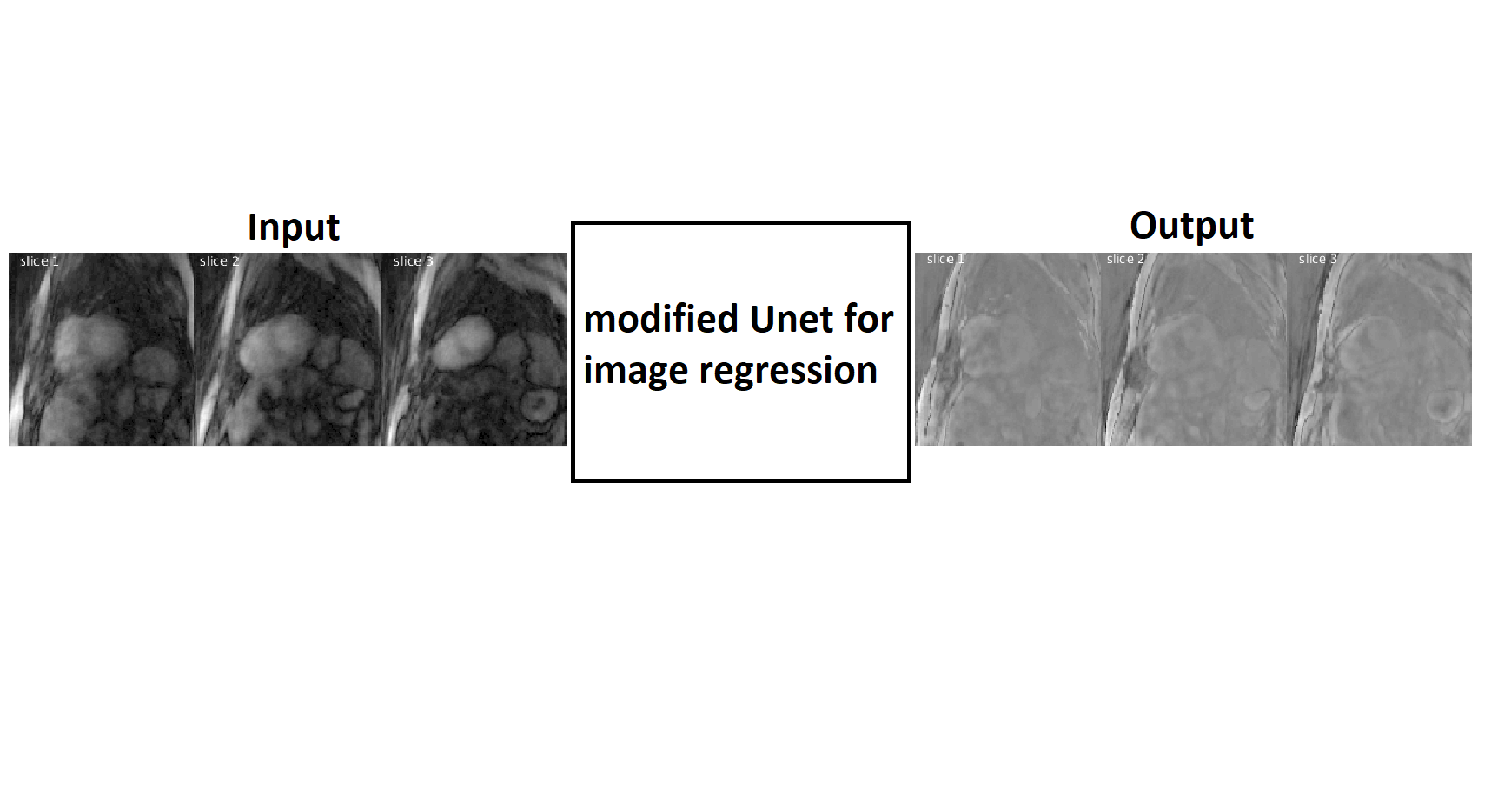
A Unet [4] convolutional neural network modified for image regression was trained on magnitude images from complex radial SMS data. This modified Unet contained 57 layers with an image input layer of 288x288x3. Three channels were used for the image input layer because each SMS set sampled three parallel slices that were reconstructed simultaneously. Ground truth images were obtained from the PT-STCR reconstruction pipeline described above. Input to the network consisted of sum of squares undersampled k-t space data reconstructed using the Inverse Fourier Transform. The network was trained to learn PT-STCR artifacts by subtracting ground-truth reconstructions from the network input. Training was done for 150 epochs with one NVIDIA K80 GPU and took ~23 hours. Figure 1 demonstrates the output and input images to the modified Unet.
Results
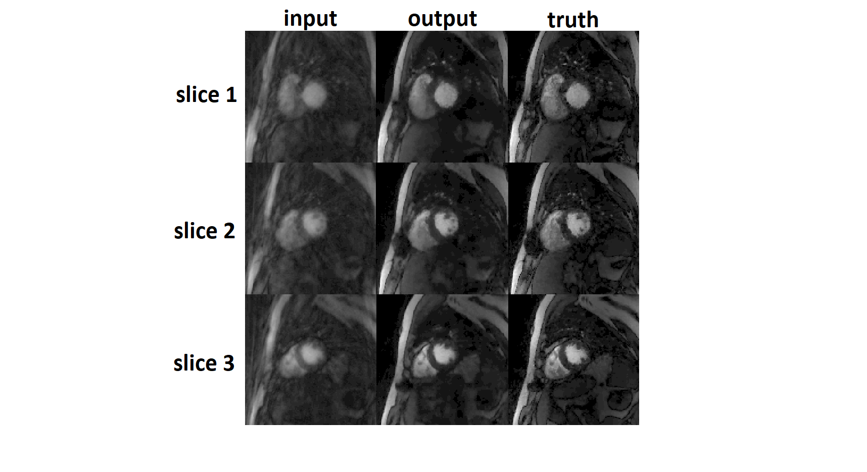
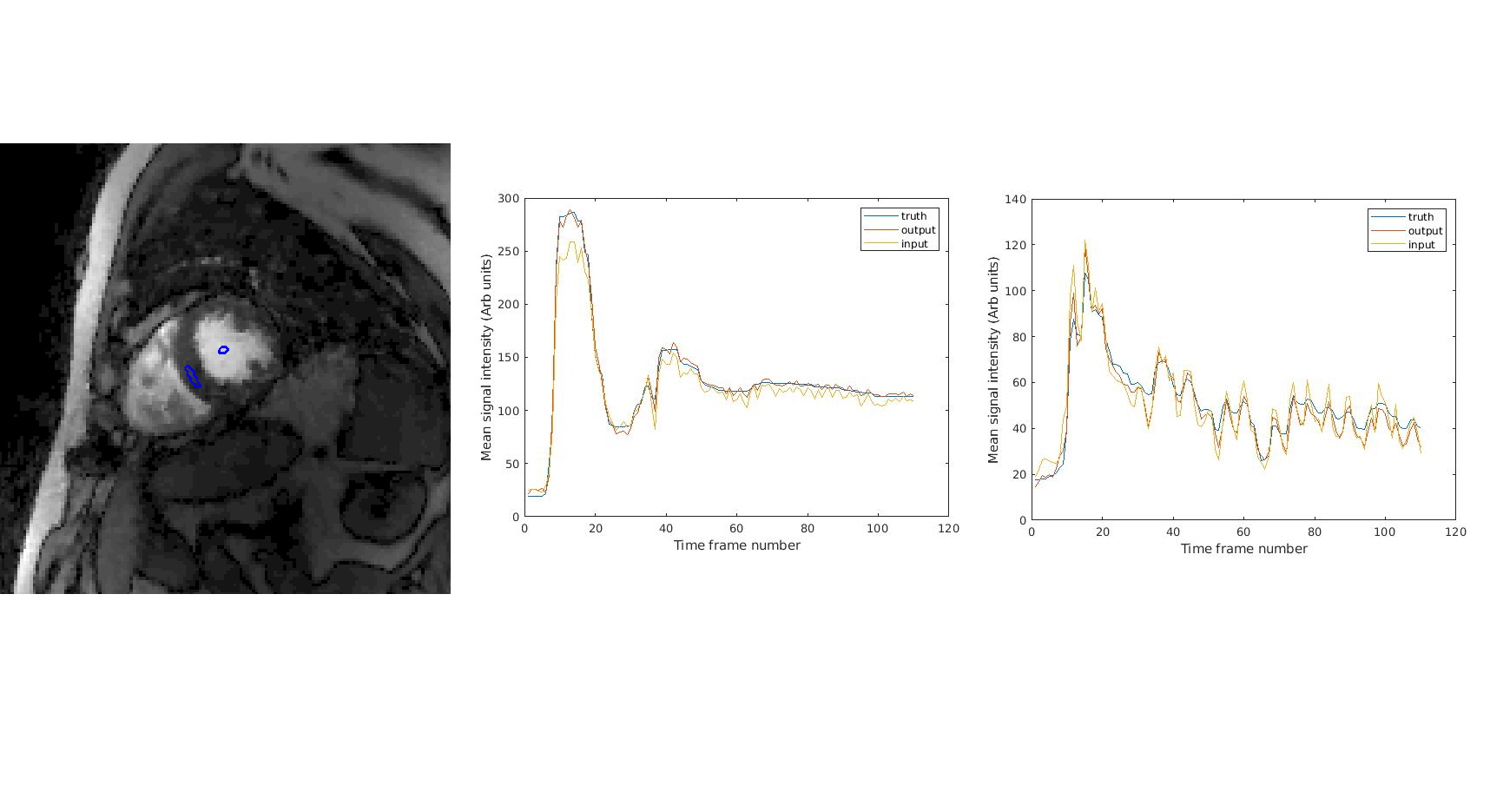
Figure 2 shows the results of the modified Unet with the residual artifact learning framework on a gated radial SMS dataset not used in training. Figure 2 demonstrates one time frame during contrast comparing the ground truth PT-STCR image with the output image from the residual artifact learning framework and the input sum of squares image for a gated radial SMS dataset. Figure 3 demonstrates the mean signal intensity time curves for regions of interest in the left ventricular blood pool and the myocardium for slice 3 of the same SMS dataset. Figure 4 demonstrates an animated gif of the same SMS dataset. This network took ~13 seconds to estimate artifacts for one SMS dataset in comparison to ~35 minutes for the PT-STCR reconstruction of the same SMS dataset.Discussion and Conclusion
Using a modified Unet for image regression coupled with a residual artifact learning framework has demonstrated promising results with improved image quality through a decrease in pixelation and blurring artifacts in comparison to the network input. This network was trained on magnitude radial SMS datasets to learn the residual artifacts as opposed to truth images as was done for image denoising [3]. These results demonstrate that high quality images can be obtained from undersampled radial SMS data for increased slice coverage and potential improvements to characterizing coronary artery diseases with a speed up factor of ~150 in comparison to the reconstruction times of PT-STCR.Acknowledgements
This research was supported by the National Heart Lung and Blood Institute of the National Institutes of Health under award number R01HL138082.References
[1] G. Adluru, J. Mendes, Y. Tian, B. Wilson, E. DiBella, Ungated myocardial perfusion imaging with complete left ventricular coverage using radial simultaneous multi-slice imaging. ISMRM 2017.
[2] G. Adluru, C. McGann, P. Speier, E.G. Kholmovski, A. Shaaban, E.V. Dibella, Acquisition and reconstruction of undersampled radial data for myocardial perfusion magnetic resonance imaging. Journal of magnetic resonance imaging : JMRI, 29 (2009) 466-473.
[3] K. Zhang, W. Zuo, Y. Chen, D. Meng, L. Zhang, Beyond a Gaussian Denoiser: Residual Learning of Deep CNN for Image Denoising, IEEE transactions on image processing : a publication of the IEEE Signal Processing Society. 26 (2017) 3142-3155
[4] O. Ronneberger, P. Fischer, T. Brox, U-Net: Convolutional Networks for Biomedical Image Segmentation. MICCAI, 9351 (2015) 234-241.
[5] E.V. Dibella, J. Mendes, M. Ibrahim, Y. Tian, B. Wilson, G. Adluru, Multiple sets of simultaneous multi-slice (SMS) for improved short and long axis coverage of myocardial DC perfusion. ISMRM 2018.
[6] Y. Tian, J. Mendes, A. Pedgaonkar, M. Ibrahim, L. Jensen, J.D. Schroeder, B. Wilson, E.V. Dibella, G. Adluru, Feasibility of multiple-view myocardial perfusion MRI using radial simultaneous multi-slice acquisitions. Manuscript submitted for publication.
[7] Y. Tian, G. Adluru, J. Mendes, E.V. Dibella, Evaluation of extended GROG and Toeplitz pre-interpolation methods on radial simultaneous multi slice MRI. ISMRM 2018.
[8] Y. Tian, A. Pedgaonkar, J. Mendes, M. Ibrahim, B. Wilson, E.V. Dibella, G. Adluru, Rapid Motion Compensation Reconstruction for Dynamic MRI using Pixel Tracking Temporal Total Variation Constraint. ISMRM 2017.
Figures