2126
Multi-Band SPIRiT Strategies for Improved Simultaneous Multi-slice Myocardial $$$T_1$$$ Mapping1Electrical and Computer Engineering, University of Minnesota, Minneapolis, MN, United States, 2Center for Magnetic Resonance Research, University of Minnesota, Minneapolis, MN, United States, 3Computer Assisted Clinical Medicine, University Medical Center Mannheim, Heidelberg University, Mannheim, Germany
Synopsis
Myocardial T1 mapping is typically performed with three or more breath-holds, each covering one slice. This leads to patient discomfort and long exam times. Recently, simultaneous multi-slice/multi-band (SMS/MB) imaging was proposed to improve coverage and reduce scan time. It was shown that k-space interpolation outperformed SENSE-type reconstructions in terms of T1 precision. However, the slice-GRAPPA approach is a kernel estimation approach and does not allow for additional regularization. In this work, we sought to use a SPIRiT approach for improving SMS/MB myocardial T1 mapping by performing regularized multi-band reconstruction.
INTRODUCTION
Myocardial T1 mapping demonstrated clinical value in numerous cardiomyopathies1. However, whole-heart coverage necessitates long scan times and multiple breath-holds, which leads to patient discomfort. For faster coverage, simultaneous multi-slice/multi-band (SMS/MB) imaging can be utilized2. A previous study has shown that accelerated SMS myocardial T1 mapping using slice-GRAPPA shows comparable accuracy with single-band imaging at the expense of reduced precision3. Since slice-GRAPPA reconstructions do not support additional regularization; SPIRiT4 is a promising candidate for further removal of artifacts arising from noise amplification and has recently been applied to SMS by enforcing self-consistency for individual slices5. In this work, we sought to develop an alternative MB-SPIRiT approach that combines the advantages of slice GRAPPA and MB-SPIRiT methods, while also allowing for regularization. This was compared to slice-GRAPPA3 and an MB-SPIRiT method5 in myocardial T1 mapping.METHODS
Sequence: Myocardial T1 mapping was performed on 5 subjects using the SAPPHIRE sequence with MB excitation of 3 slices as previously described3. FLASH imaging was used with CAIPIRINHA6 employing 2π/3 phase increments to decrease noise amplification. MB factor=3 and uniform in-plane acceleration=2 were utilized to acquire 15 different T1 weighted contrasts for 3 slices in a breath-hold with imaging parameters: FOV=320x320mm2, resolution=2x2.1mm2, slice thickness=10mm, partial Fourier = 6/8, TR/TE/FA=4/2ms/10°. Calibration data for MB imaging was acquired in a separate free-breathing scan of three slices with FOV=320x320mm2, resolution=2x5mm2, slice thickness=10mm, and TR/TE/FA=3.6/1.8ms/10°.
Slice MB-SPIRiT/GRAPPA: We propose an SMS/MB reconstruction approach to utilize advantages of both SPIRiT and slice GRAPPA using the following objective function:
$$\underset{\textbf{K}_\textbf{1},\textbf{K}_\textbf{2},\textbf{K}_\textbf{3}}{\mathrm{argmin}} \lVert P(\textbf{K}_\textbf{1}+\textbf{K}_\textbf{2}+\textbf{K}_\textbf{3})-\textbf{K}_\textbf{MB}\lVert ^2_2 + \lambda\left \lVert \textbf{G}_\textbf{MB}\textbf{K}_\textbf{MB}-\begin{bmatrix}\textbf{K}_\textbf{1}\\\textbf{K}_\textbf{2}\\\textbf{K}_\textbf{3}\end{bmatrix}\right\lVert ^2_2 + \sum_{i=1}^3\beta_i\lVert(\textbf{G}_\textbf{i}-\textbf{I})\textbf{K}_\textbf{i}\lVert^2_2 + \sum_{i=1}^3\sigma_i \lVert\psi(\textbf{K}_\textbf{i})\lVert^2_2$$
Here P is the undersampling operator in k-space, KMB is the acquired multiband k-space, GMB implements the slice-GRAPPA operation on KMB, Gi and Ki are individual coil self-consistency and individual k-space for each slice i∈{1,2,3} respectively, and $$$\psi$$$ is a regularizer. Fig. 1 depicts the schematic of data consistency (first term), Slice GRAPPA consistency (second term) and SPIRiT coil self- consistency (third term). We note that this equation reduces to the MB-SPIRiT formulation of5 when λ = 0. The objective function was solved using ADMM. Two sets of comparisons were performed. First, σi was set to 0, and slice GRAPPA, the proposed method and MB-SPIRiT were compared without regularization. Then,σi was set to 10-7 times, which is the $$$\ell_{\infty}$$$ norm of the zero filled MB image. $$$\psi(.)$$$ was selected as total variation of the image corresponding to the SENSE-1 combination of the coil images of a given slice. The proposed method with these parameters was then compared to MB-SPIRiT with the same regularization.
RESULTS
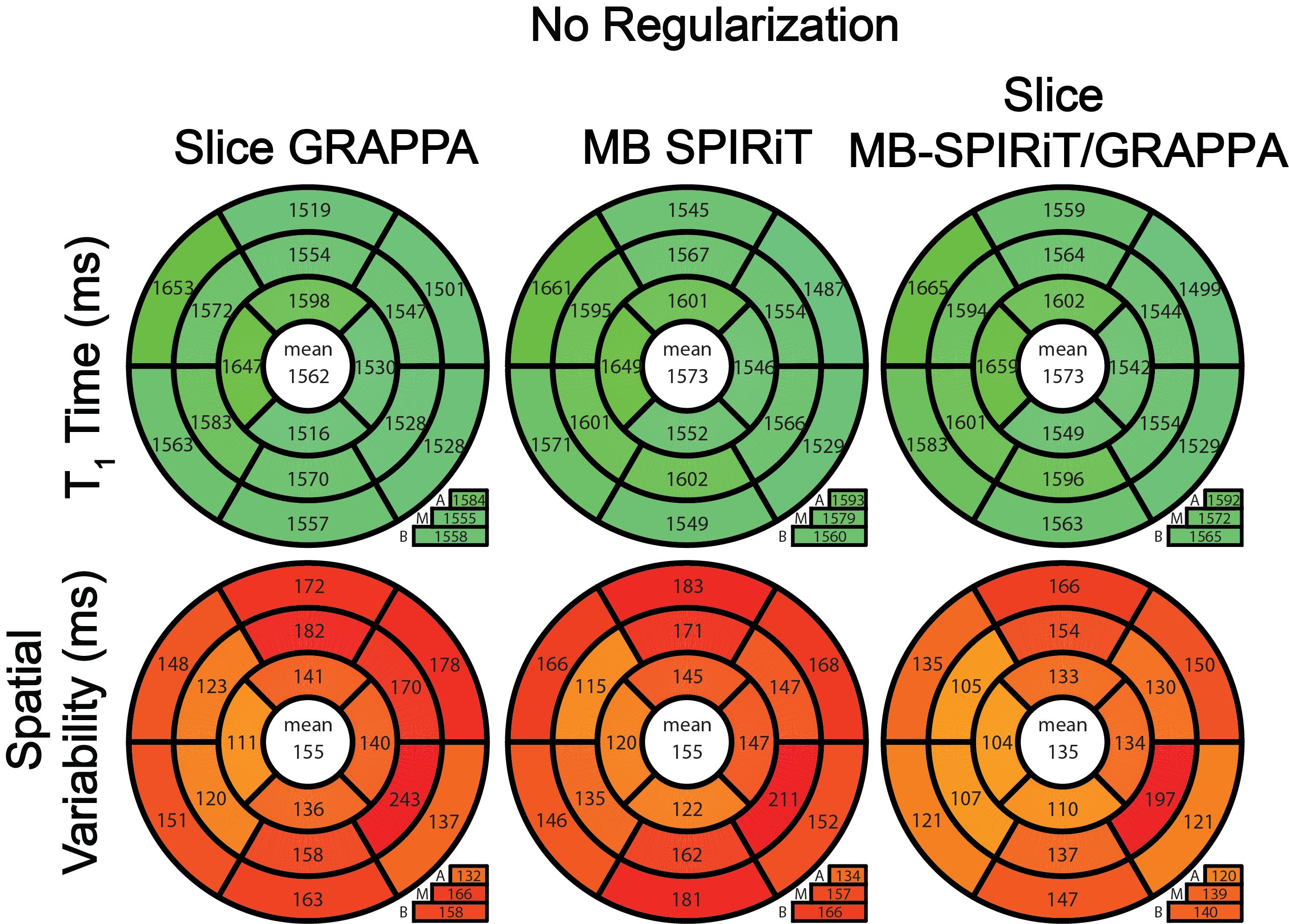
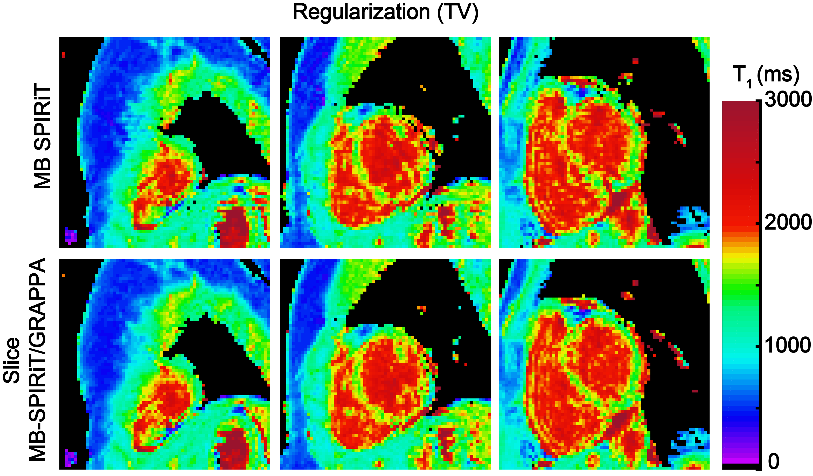
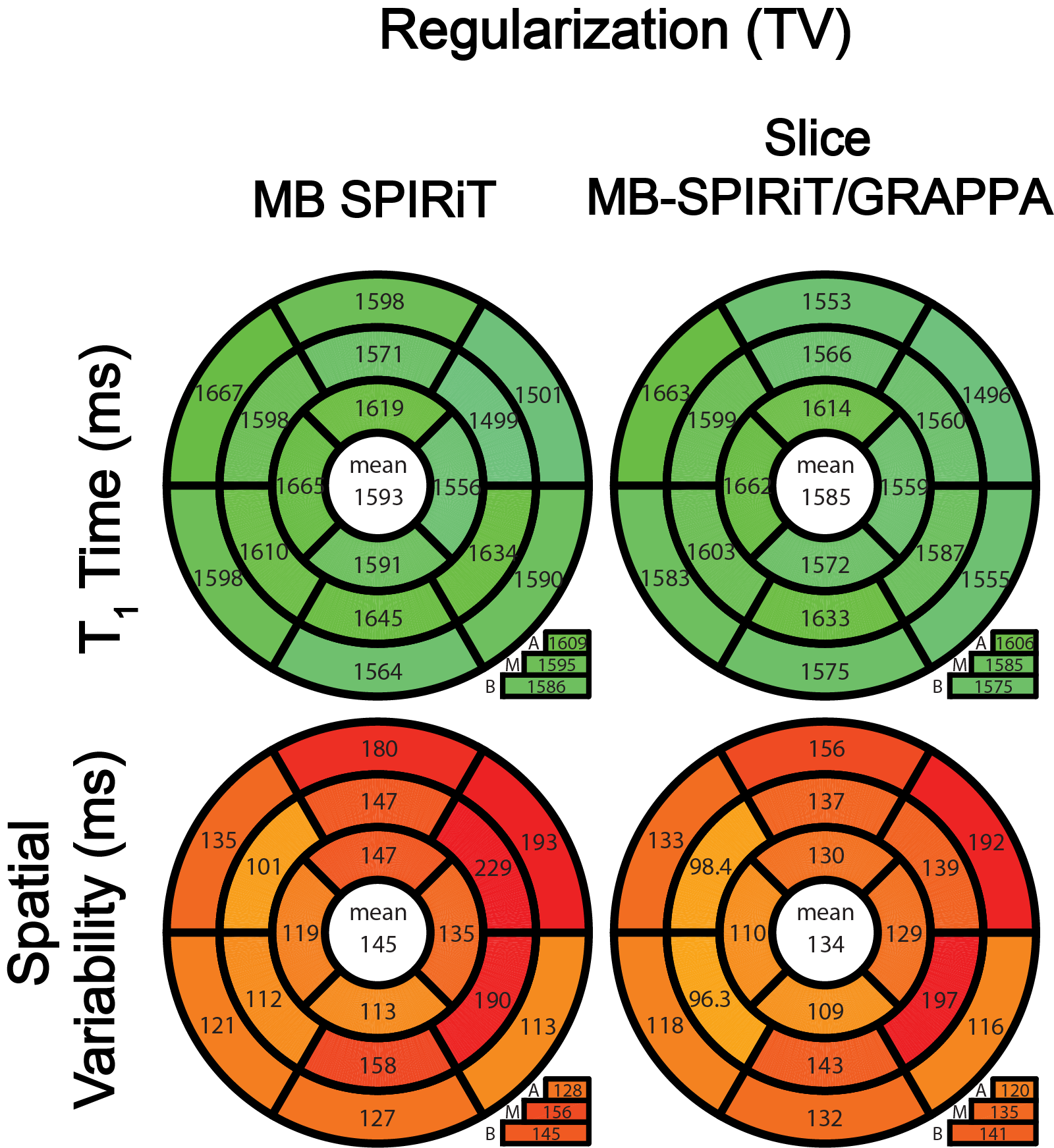
Fig. 2 shows representative MB T1 maps reconstructed without any regularization term, using slice-GRAPPA, the proposed slice MB-SPIRiT/GRAPPA, and previously described MB-SPIRiT5. Improved precision is visually observed in slice MB-SPIRiT/GRAPPA (Fig. 2). Quantitative evaluation across five subjects shows no difference among the average T1 times of the three methods. However, slice MB-SPIRiT/GRAPPA yields the least variability (135 ms), improving upon both conventional MB-SPIRiT5 and slice GRAPPA3 (both: 155 ms) (Fig. 3). T1 maps reconstructed with TV regularization using conventional MB-SPIRIT and the proposed MB-SPIRiT/GRAPPA, show visually decreased noise amplification compared with the unregularized reconstructions (Fig. 4). The proposed method with regularization also shows visually improved image quality as compared with conventional MB-SPIRIT and depicts lower spatial variation in the quantitative analysis (Fig. 5), while maintaining comparable average T1 times.DISCUSSION
In this study, we proposed and evaluated two SPIRiT based strategies for SMS/MB imaging. The proposed slice MB-SPIRiT/GRAPPA shows improved spatial variability compared to slice GRAPPA3 and MB-SPIRiT5. The SPIRiT strategies evaluated here allow for additional regularization, which may further reduce the effects of noise amplification. In this study, since our focus was on the effective use of coil information, we used a simplistic TV regularization with a very low weight as a proof of concept. More advanced regularizers5,7 may be utilized for additional benefit in practical scenarios.CONCLUSION
The proposed slice MB-SPIRiT/GRAPPA technique enables improved reconstruction of SMS-accelerated myocardial T1 mapping while allowing for incorporation of regularizers.Acknowledgements
Funding: Grant support: NIH R00HL111410, NIH P41EB015894 and NSF CAREER CCF-1651825.References
- Schelbert, Erik B., and Daniel R. Messroghli. "State of the art: clinical applications of cardiac T1 mapping." Radiology, 2016; 278(3):658-676.
- Larkman, David J., et al. "Use of multicoil arrays for separation of signal from multiple slices simultaneously excited." Journal of Magnetic Resonance Imaging, 2001; 13(2):313-317.
- Weingärtner, Sebastian, et al. "Simultaneous multislice imaging for native myocardial T1 mapping: Improved spatial coverage in a single breath‐hold." Magnetic Resonance in Medicine, 2017; 78(2):462-471.
- Lustig, Michael, and John M. Pauly. "SPIRiT: iterative self‐consistent parallel imaging reconstruction from arbitrary k‐space." Magnetic Resonance in Medicine, 2010; 64(2):457-471.
- Yang, Yang, et al. "Whole‐Heart Spiral Simultaneous Multi‐Slice First‐Pass Myocardial Perfusion Imaging." Magnetic Resonance in Medicine, in press; doi:10.1002/mrm.27412
- Breuer, Felix A., et al. "Controlled aliasing in parallel imaging results in higher acceleration (CAIPIRINHA) for multi‐slice imaging." Magnetic Resonance in Medicine, 2005; 53(3):684-691.
- Zhang, Tao, John M. Pauly, and Ives R. Levesque. "Accelerating parameter mapping with a locally low rank constraint." Magnetic Resonance in Medicine (2015); 73(2):655-661.
Figures
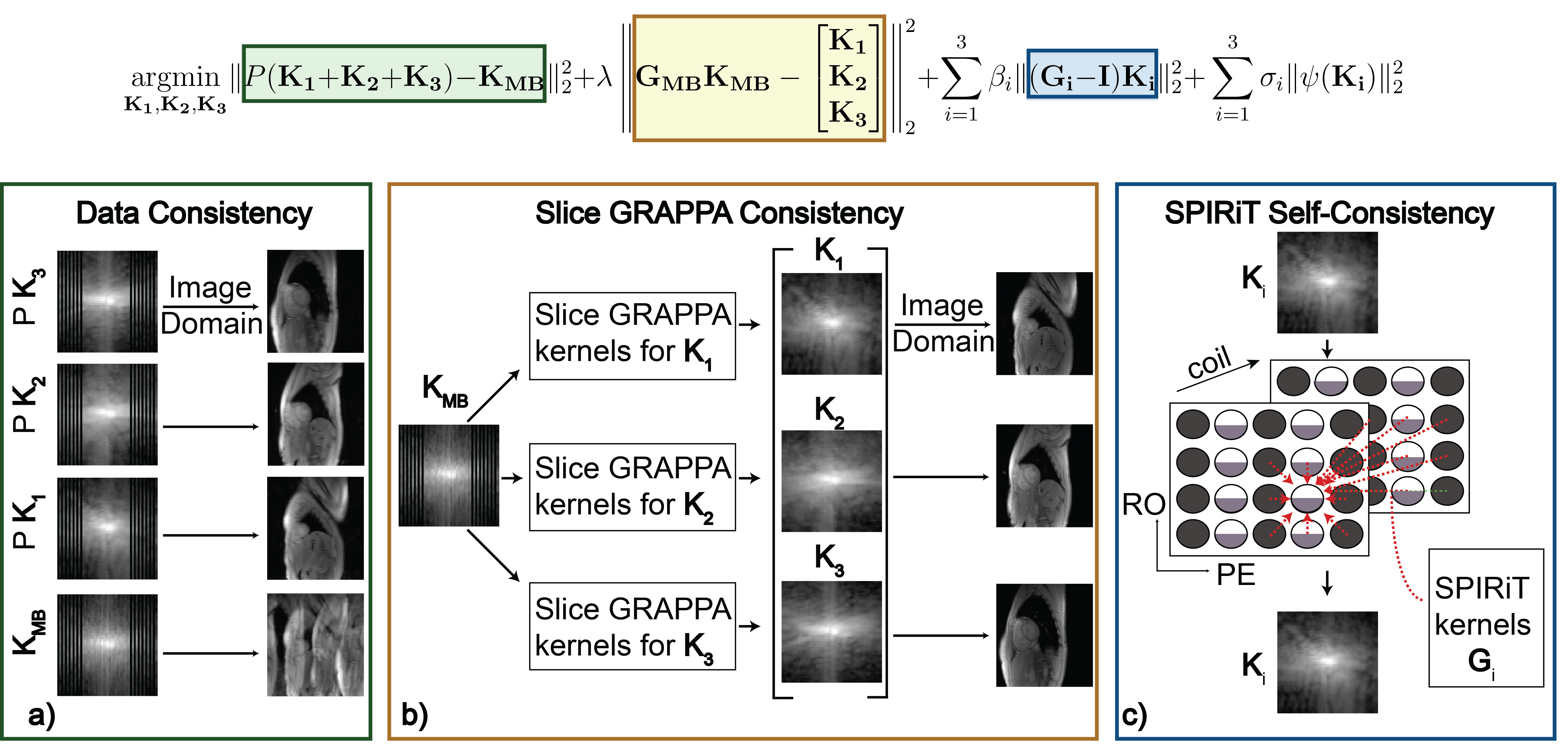
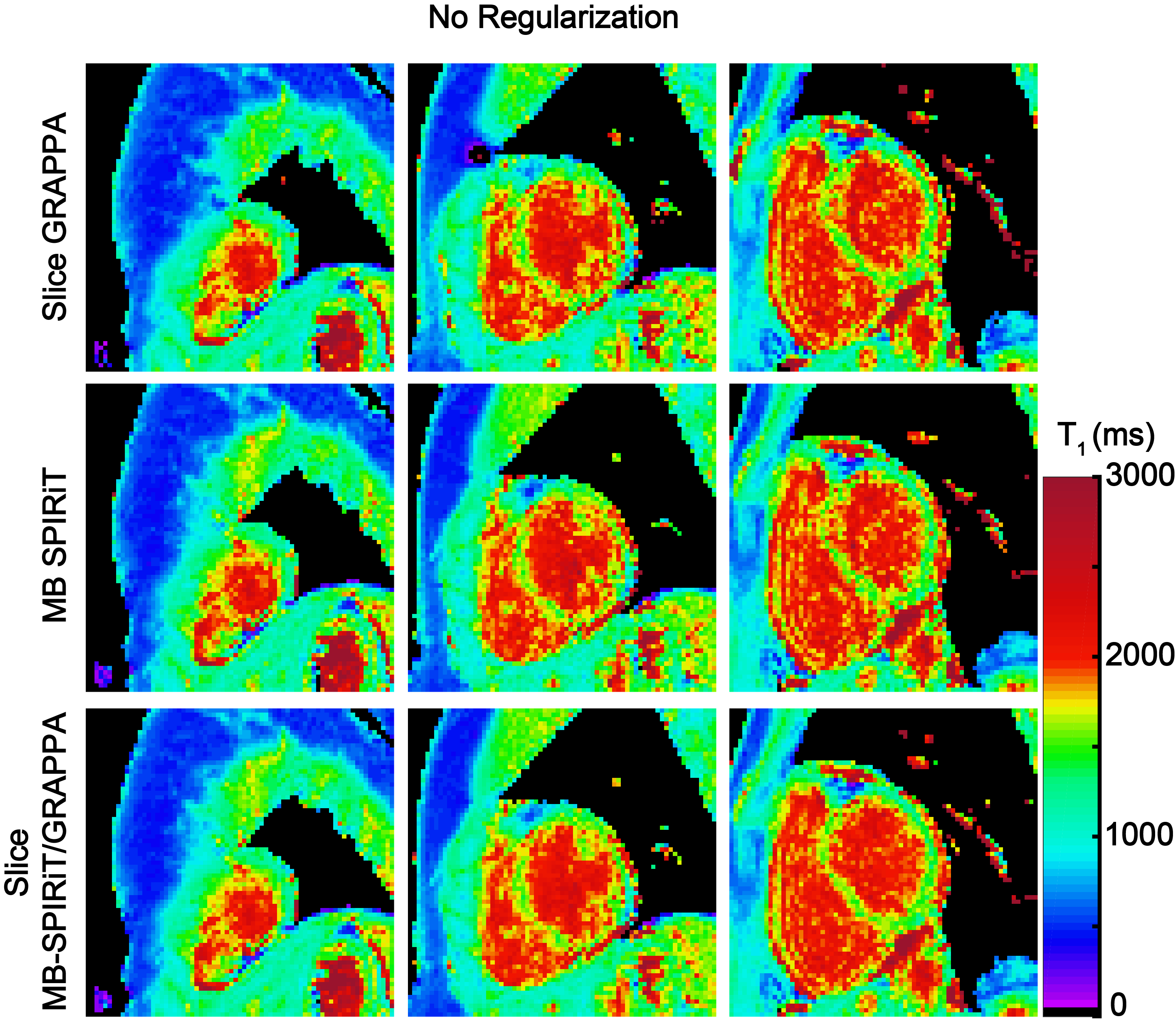
Figure 2: Representative T1 maps comparing with slice GRAPPA3, MB-SPIRiT5 and slice MB-SPIRiT/GRAPPA. Slice GRAPPA images were obtained with [5,5] slice-GRAPPA kernels followed by [5,4] in-plane GRAPPA kernels respectively3. MB-SPIRiT was implemented with the following parameters were used λ=0, β 1 = β2 = β3 = 10-3 and σ1 = σ2 = σ3 = 0. Slice MB-SPIRiT/GRAPPA images were reconstructed with the above parameters for β i and σ i, but with λ = 10-4. All SPIRiT reconstructions were calibrated using [7,7] kernels. MB-SPIRiT/GRAPPA depicts visually highest image quality with the best homogeneity throughout the myocardium.