2105
Tiny Golden Angle Real-time Cardiac MRI in the Mouse Model1Core facility Small Animal Imaging, Ulm University, Ulm, Germany, 2Department of Cardiology, The Second Hospital of Shandong University, Jinan, China, 3Department of Internal Medicine II, Ulm University Medical Center, Ulm, Germany
Synopsis
Even though an established imaging method, small animal cardiac MRI is a time-consuming technique. Where self-gating techniques enabled wider-spread applications, acquisition times in the minute range for a single slice still limit its application for physiological stress or first-pass perfusion imaging. We translated the tiny golden angle sparse sense approach to small animal application. The technique was tested in ten animals and the resulting functional parameters compared with the self-gating approach. Further, initial application for pharmacological stress imaging was tested in three animals.
Purpose
To investigate the feasibility of tiny golden angle radial sparse MRI for real-time imaging of cardiac function in the mouse model.Introduction
The small size of the mouse heart (approximately 1/2000th the mass of a human heart), high heart rates (about 250–650 beats per minute, depending on the depth of anaesthesia), and high respiratory rates (about 60–160 cycles per minute (cpm)) impose substantial challenges for functional assessment by MRI. Conventional ECG-gated or triggered approaches face severe limitations due to the cumbersome ECG recording, which is often of insufficient quality especially at high field strengths and during continuous scanning due to gradient induced signal distortion. Over the last years, self-gating techniques (1) have been introduced enabling high-quality cardiac MRI with high reproducibility (2,3). To reduce acquisition times application of real-time methods have been suggested and initially evaluated (4). Tiny golden angle sparse sense (tyGRASP) has proven beneficial artefact properties for real-time cardiac imaging at 3T (5). In this contribution, the tyGRASP approach was evaluated for assessment of cardiac function in mice at 11.7T.
Methods
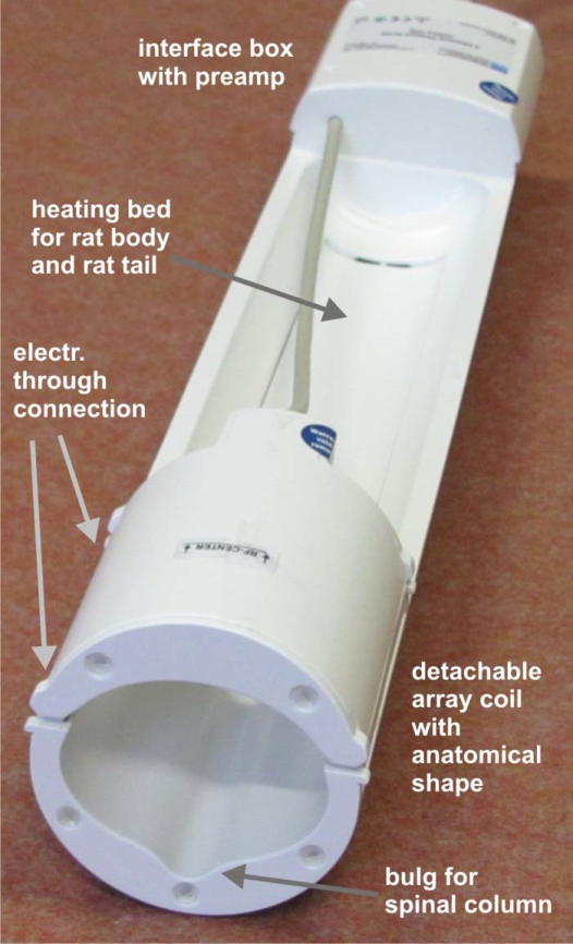
A tiny golden angle trajectory was implemented on a small animal MRI system (BioSpec 117/16, Bruker Biospin, Ettlingen, Germany). Prior to each acquisition the trajectory was mapped along the three spatial dimensions to allow for compensation of trajectory distortions during reconstruction. All data were acquired with a four-element thorax coil (Fig. 1, RAPID Biomedical, Rimpar, Germany). A tiny golden angle of φ7 =23.62814° with G37=8 and G47=15 projections for homogenous k-space coverage. Further acquisition parameters include: TE = 860µs, TR = 2.1ms,TACQ=1.2s slice, FOV = 3x3cm², and 128 readout samples yielding Δr=0.2342mm2 resolution. For minimizing TE, an asymmetric sinc-gauss excitation pulse with 1.5kHz BW was used. For improving blood-myocardium contrast a rather high flip angle α=20° was chosen. Self-gated data were acquired as reference with a high-resolution (IG-HR, TR/TE = 1.0/5.75ms,Δr=0.1172 mm2 ,TACQ=2m30s per slice) and a low-resolution (IG-LR TR/TE = 1.0/5.75ms,Δr=0.22 mm2, TACQ=42s per slice) protocol. The tyGRASP data were reconstructed with in-house build reconstruction framework, implemented in Matlab (Matlab,The MathWorks,Natick, Massachusetts,USA), applying a compressed sense reconstruction with total variation sparsity operator (5) using G37=8 (RT1) and G47=15 (RT2) projections for the reconstruction of a single time frame, yielding a temporal resolution of 16.8ms (RT1) and 31.5ms (RT2).
The imaging protocol was performed in 10 wildtype (C57/B6) mice for a short axis stack completely covering the left ventricle. During scanning the animals were kept at mild anaesthesia (cpm > 60). End-diastolic (EDV), end-systolic (ESV), stroke volume (SV), and ejection fraction (EF) were quantified with Segment (Medviso AB, Lund, Sweden) and compared between RT2 and IG-HR different acquisition protocols. Statistical significance of the differences was assessed applying a two-tailed paired T-test, with p-values below 0.05 being considered significant.
Three mice were additionally scanned after injection of 1.5µg/g bodyweight dobutamine for initial evaluation of the real-time techniques under physiological stress.
Results
The acquisition protocol could be completed in all mice. Two mice had to be excluded due to severe image artefacts in all imaging protocol most likely caused by animal motion during scanning due to the only mild anaesthesia. Resulting image quality for the different imaging protocols is provided in Fig. 2. Even though the non-cardiac structures are not clearly visible in the real-time images, the vascular structures as well as the left ventricle can be well appreciated and quantitative assessment of the functional parameters was possible in the analysed real-time data with no significant differences for rest (Fig. 3). For the stress investigation, a trend to underestimation of the volumes by the real-time approach can be observed.Conclusion
Real-time cardiac tyGRASP in mice appears feasible with sufficient image quality for quantification of at least global functional parameters. From as little as G37=8 projections (Δt=16.8ms) acceptable image quality for assessment of the cardiac function could be obtained. Image quality clearly improved when using G47 = 15 projections (Δt=31.5ms). Even under pharmacological stress, sufficient image quality for functional analysis could be achieved. Whether the slight underestimation of the volumes in real-time imaging is due to the different time-points of the acquisition relative to drug administration or due to the limited spatial resolution of the real-time method needs further investigation. The tyGRASP approach enables flexible number of projections for image reconstruction thus offering the possibilty for cardiac-phase dependent adjustment of the temporal resolution, which might further optimize the resulting image quality. With the current image quality, new applications like first-pass perfusion imaging may be addressed in the future.
Acknowledgements
No acknowledgement found.References
1) Hiba B, Richard N, Janier M, Croisille P. Cardiac and respiratory double self-gated cine MRI in the mouse at 7T. Magn Reson Med 2006; 55:506-513.
2) Joubert M, Tager P, Legallois D, Defourneaux E, Le Guellec B, Gerber B, Morello R, Manrique A. Test-retest reproducibility of cardiac magnetic resonance imaging in healthy mice at 7-Tesla: effect of anesthetic procedures. Sci Rep. 2017 Jul 27;7(1):6698.
3) Zuo Z, Subgang A, Abaei A, Rottbauer W, Stiller D, Ma G, Rasche V. Assessment of Longitudinal Reproducibility of Mice LV Function Parameters at 11.7 T Derived from Self-Gated CINE MRI. Biomed Res Int. 2017;2017:8392952.
4) Wech T, Seiberlich N, Schindele A, Grau V, Diffley L, Gyngell ML, Borzì A, Köstler H, Schneider JE. Development of Real-Time Magnetic Resonance Imaging of Mouse Hearts at 9.4 Tesla--Simulations and First Application. IEEE Trans Med Imaging. 2016 Mar;35(3):912-20.
5) Wundrak S, Paul J, Ulrici J, Hell E, Geibel MA, Bernhardt P, Rottbauer W, Rasche V. Golden ratio sparse MRI using tiny golden angles. Magn Reson Med. 2016 Jun;75(6):2372-8.
Figures