2096
A fast and contrast-free MR approach to the diagnosis of deep vein thrombosis based on DANTE-prepared gradient echo1School of Basic Sciences, Guangzhou Medical University, Guangzhou, China, 2Department of Radiology, Nanshan people’s hospital, Shenzhen, China, 3Department of Radiology, Panyu Central hospital, Guangzhou, China, 4MR Collaborations, Siemens Healthcare Ltd, Shenzhen, China, 5MR R&D, Siemens Healthineers, Los Angeles, CA, United States, 6Paul C. Lauterbur Biomedical Imaging Center, Shenzhen Institutes of Advanced Technology, Shenzhen, China, 7Biomedical Imaging Research Institute, Cedars Sinai Medical Center, Los Angeles, CA, United States
Synopsis
Black-blood magnetic resonance thrombus imaging (BTI) has proved successful for the diagnosis of deep vein thrombosis (DVT) with high sensitivity, specificity, and accuracy. Previous BTI method is based on a 3D variable flip angle fast spin echo (3D-vFSE) sequence, which has high specific absorption rate (SAR). DANTE-FLASH is another black-blood MR technique previously proposed for vessel wall imaging at lower extremities1. We hypothesized that DANTE-FLASH may suffice the purpose of diagnosing DVT while avoiding high SAR. This work was aimed to investigate the feasibility of using DANTE-FLASH to diagnose DVT.
PURPOSE:
Deep vein thrombosis
(DVT) has an estimated annual incidence of approximately 5 per 10,000 in the
general population2. DVT can be diagnosed by contrast-free magnetic resonance (MR) imaging,
such as MR direct thrombus imaging (MRDTI)3 and black-blood MR thrombus imaging (BTI)4. Unlike MRDTI that relies on the short-T1 methemoglobin within the subacute
thrombus to produce high signals on T1-weighted images, BTI can directly
visualize the thrombus within the venous lumen by suppressing the venous blood
flow signal 4, 5. DANTE-FLASH is another black-blood MR technique previously proposed for
vessel wall imaging at lower extremities1. Compared to BTI, which is based on a DANTE-prepared 3D fast spin-echo
sequence, DANTE-FLASH may suffice the purpose of diagnosing DVT while avoiding high
specific absorption rate. This work was aimed to investigate the feasibility of
using DANTE-FLASH to diagnose DVT.
METHODS:
Experiments
The IRB approved study was performed on a 3 T scanner (Skyra, Siemens Healthcare, Erlangen, Germany), and consecutively enrolled 6 healthy volunteers (6 M, aged 22–48 years) and 19 patients (13 M,6 F, aged 28–90 years). All subjects underwent three-station DANTE-FLASH scan to cover bilateral lower limbs. Ultrasound and MR direct thrombus imaging (MRDTI) was also conducted to provide a standard reference. The scan parameters for the DANTE module included: flip angle = 15˚, pulse train length =150. The parameters for the FLASH readout included: echo spacing/TE = 5.4/2.08 ms, flip angle =12˚, isotropic resolution = 1.2×1.2×1.2 mm3 and reconstructed to 0.6×0.6×0.6 mm3, FOV = 380×380 mm2, bandwidth=425 Hz/pixel, parallel imaging (GRAPPA) factor = 2, partial Fourier factor = 7/8, TR =800 ms, water excitation for fat suppression. The scan parameters of MRDTI were the same as those of FLASH readout in DANTE-FLASH except the inversion recovery time (200ms) and flip angle (18 ˚).
Image Analysis
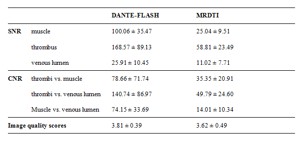
All MR images were loaded to a workstation (Leonardo; Siemens AG, Germany) for review and analysis. To quantitatively analyze the image quality of DANTE-FLASH and MRDTI, image signal-to-noise ratio (SNR) ,apparent contrast to noise ratio (CNR) between muscle and dark venous lumen w, and CNR between thrombus and dark venous lumen . To qualitatively analyze the diagnosis performance of DANT-FLASH, two radiologists independently assessed the randomized images and made a diagnosis of DVT on both DANTE-FLASH and MRDTI. Image quality was scored was rated by one radiologist independently on a 4-point scale (1 = poor, 4 = excellent). The sensitivity (SE), specificity (SP), positive and negative predictive values (PPV and NPV), and accuracy (ACC) of DANTE-FLASH were then calculated. Clot burden was also scored by the two radiologists in consensus according to previous study6: 0, patent vein segment; 1, nonocclusive thrombus; 2, subsegmental, occlusive thrombus; and 3, occlusive thrombus of the entire length of segment. Clot burden was compared between DANTE-FLASH and MRDTI . The Wilcoxon signed rank test was used to determine the differences of clot burden scores. A p value of less than 0.05 was considered statistically significant.
RESULTS:
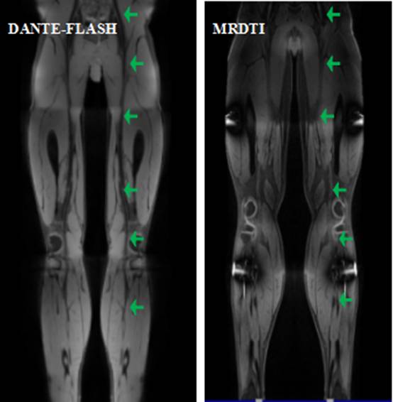
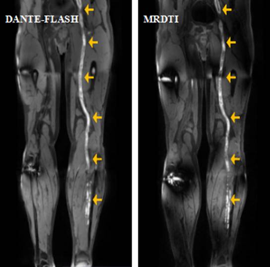
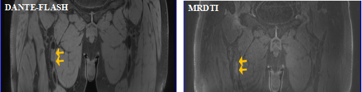
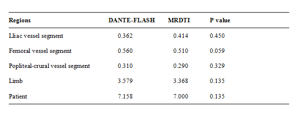
The MR and US scans were successfully conducted without any adverse events. Compared to MRDTI, DANTE-FLASH achieved better venous lumen delineation (Figure 1) and better image quality in terms of SNR, CNR, and image quality scores (Table 1). Thrombi were correctly identified by DANTE-FLASH and matched well with those detected by MRDTI (Figure 2). It was noted by radiologists that the iso-intense thrombus was easier to identify using DANTE-FLASH method as compared to that images acquired from MRDTI sequence (Figure 3). Using US as standard reference, the diagnosis SE, SP, PPV, NPV and ACC of DANTE-FLASH were 76.10%, 91.51%, 75.00%, 97.00%, and 91.18%, respectively. Using MRDTI as standard reference, the diagnosis SE, SP, PPV, NPV and ACC of DANTE-FLASH were 92.31%, 100%, 100%, and 97.00%, respectively. The clot burden obtained with DANTE-FLASH was not significantly different from MRDTI based on region, limb, or patient (Table 2).CONCLUSION:
DANTE-FLASH adequately suppresses venous blood signals and allows for direct visualization of thrombus even if the signal of the thrombus signal is iso-intense. It may serve as a safe and convenient alternative for the diagnosis of DVT.Acknowledgements
No acknowledgement found.References
1. Xie G, Zhang N, Xie Y, et al. DANTE-prepared three-dimensional FLASH: A fast isotropic-resolution MR approach to morphological evaluation of the peripheral arterial wall at 3 Tesla. J Magn Reson Imaging 2016;43(2):343-51.
2. Fowkes FJI, Price JF, Fowkes FGR. Incidence of Diagnosed Deep Vein Thrombosis in the General Population: Systematic Review. European Journal of Vascular & Endovascular Surgery 2003;25(1):1-5.
3. Moody AR. Magnetic resonance direct thrombus imaging. Journal of Thrombosis & Haemostasis 2010;1(7):1403-9.
4. Xie G, Chen H, He X, et al. Black-blood thrombus imaging (BTI): a contrast-free cardiovascular magnetic resonance approach for the diagnosis of non-acute deep vein thrombosis. J Cardiovasc Magn R 2017;19(1).
5. Chen H, He X, Xie G, et al. Cardiovascular magnetic resonance black-blood thrombus imaging for the diagnosis of acute deep vein thrombosis at 1.5 Tesla. J Cardiovasc Magn R 2018;20(1).
6. Protack CD, Bakken AM, Patel N, et al. Long-term outcomes of catheter directed thrombolysis for lower extremity deep venous thrombosis without prophylactic inferior vena cava filter placement. J Vasc Surg 2007;45(5):992-7.
Figures