2089
Irregularity of Carotid Atherosclerotic Plaque Determined by Multidimensional Magnetic Resonance Imaging Stratifies the Risk of Acute Cerebral Infarction1Department of Radiology, Nanjing BenQ Hospital, Nanjing, China, 2Nanjing Medical University, Nanjing, China, 3Center for Biomedical Imaging research, Department of Biomedical Engineering, Tsinghua University School of Medicine, Beijing, China
Synopsis
Carotid plaque surface features including surface irregularities and ulceration represent an important indicator for plaque vulnerability and are associated with the occurrence of neurological symptoms1-2. This study aimed to determine the association between surface irregularity of carotid plaque on MR images at multi-dimensions and acute ischemic stroke. Ninety-seven symptomatic patients with atherosclerotic plaque in carotid artery determined by ultrasound imaging were enrolled and underwent MR imaging for carotid artery and brain. We found that the irregularity information at multi-dimensions which combined the axial and oblique images had stronger predictive value for acute ischemic lesion than axial images alone.
Introduction and purpose
Carotid plaque surface features including surface irregularities and ulceration represent an important indicator for plaque vulnerability and are associated with the occurrence of neurological symptoms1-2. Accurate identification of plaque surface irregularity is, therefore, important for stratification of the risk of cerebral ischemic events. Magnetic resonance (MR) vessel wall imaging has been largely utilized to characterize carotid plaque features with excellent reliability and accuracy. However, the previous approach in assessing carotid plaque surface characteristics is based on single-dimension information derived from either axial MR images or oblique images3-4. We hypothesized that multi-dimensions information by combining axial and oblique MR images may have incremental value in stratifying risk of ischemic stroke. This study sought to determine the association between surface irregularity of carotid plaque on MR images at multi-dimensions and acute ischemic stroke.Methods
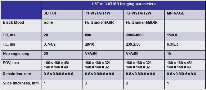
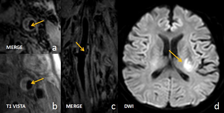
Study sample: Ninety-seven symptomatic patients (mean age, 54.7±15.4 years; 66 males) with atherosclerotic plaque in at least one carotid artery determined by B-mode ultrasound imaging (intima-media thickness ≥1.5 mm) were enrolled and underwent MR imaging for carotid artery and brain. MR imaging: The MR imaging was performed on 1.5 or 3.0T Philips MR scanners with 8-channel head and carotid coils. The multicontrast MR imaging protocol for carotid artery was detailed in Table 1. We also acquired 3D T1-VISTA /sequence obliquely at 1.5T and 3D MERGE sequence (GR; TR/TE 9.3/4.4ms; flip angle 6; spatial resolution 0.8×0.8×0.8 mm3) coronally at 3.0T for carotid artery bifurcation, respectively. A routine protocol was used to acquire T1W, T2-FLAIR and DWI sequences for brain imaging. Image analysis: The MR images of symptomatic carotid arteries were analyzed to determine the irregularity of plaque in two separate rounds of review. In the fist round of review, the presence or absence of irregular surface of carotid plaque was identified by 2 reviewers with consensus on the multicontrast MR axial images (single-dimension) blinded to oblique MR images and clinical information. After one month of time interval, in the second round of review, the 2 reviewers determined the irregularity of plaques on both multicontrast axial images and oblique T1-VISTA or multiplanar project reconstructed (MPR) images of 3D MERGE (multi-dimensions) with consensus blinded to clinical information. Presence/absence of acute ischemic lesion (AIL) on DWI images was also assessed. Statistical analysis: The correlation between irregularity of carotid plaque surface and AIL was analyzed using logistic regression and receiver-operating-characteristic curve (ROC).Results
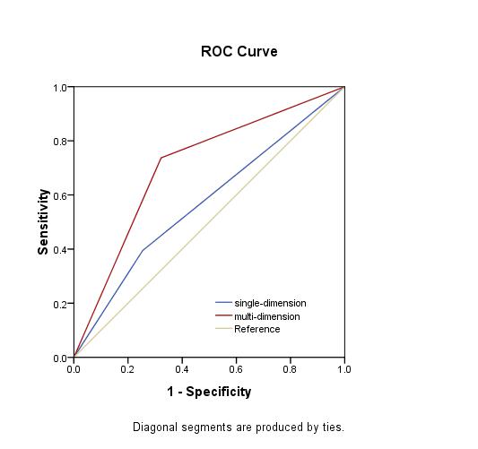
Of 97 patients, 38 (39.2%) had AILs. On MR images at single-dimension (axial MR images), 30 (30.9%) plaques were found to have irregular surface. In contrast, on the MR images at multi-dimensions (both axial and oblique MR images), 47 (48.5%) plaques were found to have irregular surface. The odds ratio (OR) of irregularity of plaque surface determined by MR images at single-dimension and multi-dimensions was 1.91 (95% CI 0.80-4.59, p =0.146) and 5.90 (95% CI 2.38-14.58, p <0.001) in discriminating presence of AIL, respectively. After adjusted for demographic characteristics and traditional risk factors, the OR of single-dimension and multi-dimensions was 2.38 (95% CI 0.92-6.17, p =0.075) and 10.31 (95% CI 3.37-31.60, p <0.001), respectively. ROC curves showed that, in discriminating presence of AIL, the area-under-the-curve (AUC) of irregularity of plaque surface determined by MR images at single-dimension and multi-dimensions was 0.57 (95% CI 0.45-0.69, p =0.244) and 0.71 (95% CI 0.60-0.81, p =0.001), respectively.Discussion and Conclusion
This study investigated the usefulness of the irregularity of plaque surface determined by MR imaging at multi-dimensions in stratifying risk of acute ischemic stroke. We found that the irregularity of plaque surface determined by MR imaging at multi-dimensions was independently associated with acute ischemic stroke. We also found that the irregularity information at multi-dimensions which combined the axial and oblique imaging had stronger predictive value for acute ischemic lesion than axial imaging alone.Acknowledgements
None.References
1. Saba L, Anzidei M, Sanfilippo R, Montisci R, Lucatelli P, Catalano C, Passariello R, Mallarini G. Imaging of the carotid artery. Atherosclerosis. 2012;220(2):294-309.
2. Singh TD, Kramer CL, Mandrekar J, Lanzino G, Rabinstein AA. Asymptomatic Carotid Stenosis: Risk of Progression and Development of Symptoms. Cerebrovasc Dis. 2015;40(5-6):236-243.
3. Yu W, Underhill HR, Ferguson MS, Hippe DS, Hatsukami TS, Yuan C, Chu B. The added value of longitudinal black-blood cardiovascular magnetic resonance angiography in the cross sectional identification of carotid atherosclerotic ulceration. J Cardiovasc Magn Reson. 2009;11(1):31.
4. Troyer A, Saloner D, Pan XM, Velez P, Rapp JH, et al. Major carotid plaque surface irregularities correlate with neurologic symptoms. J Vasc Surg. 2002;35(4):741-747.
Figures