2087
Carotid Artery Self-navigation Coughing Motion Correction Using Principle Component Analysis Jointed with Differential Threshold Method1Department of Biomedical Engineering, School of Medicine, Tsinghua University, Beijing, China, 2School of Biomedical Engineering and Imaging Sciences, King's College London, London, United Kingdom
Synopsis
Black-blood MRI is an important tool to evaluate atherosclerosis for carotid artery, however, it is susceptible to motion and no former motion correction method for carotid artery could achieve 100% acquisition efficiency. Therefore, a 100% acquisition efficiency self-navigation carotid artery motion correction method based on Principle Component Analysis (PCA) jointed with Differential Threshold Method (DTM) was proposed for carotid artery imaging using radial sampling. Experimental results demonstrated that the proposed method can detect acute motions such as coughing, compute the motion-corrupted interval, and achieve higher reconstruction quality after removing the motion-corrupted spokes.
Introduction
During the scan of carotid artery wall imaging, motions will blur the image and make it hard for doctors to diagnose1. Detection and removal of these motions, especially acute motions such as coughing and swallowing, are important to improve image quality. Common motion correction methods for carotid artery include navigator echoes2, structured light3 or pre-scan1, all of which have the drawbacks of either low data acquisition efficiency or auxiliary hardware-based gating, resulting prolonged total scan time or complicated scan procedure. Since the center of k-space data has the potential for self-navigation, this work aimed to proposed a 100% acquisition efficiency self-navigation approach using PCA jointed with DTM4 to detect motion occurrence and remove motion-corrupted spokes by analyzing radial sampling k-space center data.Methods
Data Acquisition: A total of 31 datasets were required from 6 free-breathing healthy volunteers on a 3T scanner (Philips, Achieva) with 8-channel carotid coil, using a 3D Golden-Angle Radial Stack-of-Stars SPGR sequence5. The scan parameters were TR=7.7ms, TE=3.4ms, flip angle=15°, FOV=160×160×60 mm3 with 0.8×0.8 mm2 in-plane resolution and 2 mm slice thickness. Because coughing accompanied fierce abdominal move, a bellow recording the abdomen motion would serve as the ground truth. Volunteers were instructed to randomly cough 1 to 3 times during the scan.
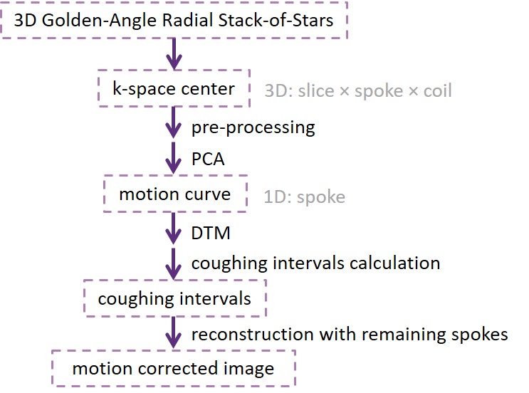
Theory: The pipeline for the whole procedure was demonstrated in Fig. 1. A motion curve representing moving distance along time was acquired via employing PCA and coil clustering to the center of k-space data6,7. To be exact, after applying PCA along slice dimension for each coil, coil clustering was performed to the concatenated large matrix to selected good coils, which were then be integrated into a 2D matrix. Another PCA would be done along slice jointed coil dimension and regard the first principle component as the final motion curve. Note that filtering with hamming window and no normalization before PCA can achieve better results. Because carotid artery motion will inevitably include respiratory and cardiac motions, DTM was used to amplify and select desired relatively acute coughing peaks: 1. applying a low-pass filter: reduce high-frequency interference such as respiratory or cardiac motions; 2. differential and squaring operators: strengthen coughing peaks and weaken small-amplitude signals; 3. smoothing: use discrete convolution to smooth the signal such that there is only one peak in one coughing occurrence. Based on the characteristic of coughing pattern, an interval will be automatically calculated and returned for each coughing peak. The spokes in these intervals will be removed in the reconstruction. Remained spokes were reconstructed with iGRASP8.
Results
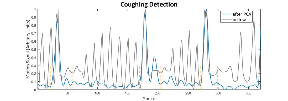
Fig. 2 demonstrates the motion signal processed by PCA and automatically calculated coughing intervals with the reference of bellow. We can see that coughing intervals correspond perfectly to coughing peaks of bellow motion curve, proving that this algorithm can correctly detect coughing occurrence. For 31 datasets, there are a total number of 58 coughs according to the indication of bellow and the instruction recording during the scan, with 57 coughs correctly detected (True Positive Value: 98.3%) and 7 coughs redundantly detected (False Positive Value: 12.1%). After removing the spokes in coughing intervals returned by this proposed algorithm, reconstruction image quality has been improved than original images with carotid artery walls much clearer and continuous in Fig. 3.
Discussion and Conclusions
This result demonstrates that the proposed PCA jointed with DTM can accurately detect coughing occurrence with True Positive Value of 98.3% for all subjects, suggesting this method is feasible for motion correction in carotid artery imaging. The limitations in this work may be the absence of direct carotid artery motion indicator because bellow is an indirect signal for carotid artery and cannot record the occurrence of swallowing or neck moving. Therefore, the redundant detections of coughs may result from the existence of those non-recorded motions.
In conclusion, a retrospective self-navigation approach has been developed to detect acute motion during carotid artery MR imaging. Its effectiveness and robustness in reducing coughing-related motion artifacts were validated.
Acknowledgements
No acknowledgements found.References
[1] Fan Z, Zuehlsdorff S, Liu X, et al. Prospective self‐gating for swallowing motion: A feasibility study in carotid artery wall MRI using three‐dimensional variable‐flip‐angle turbo spin‐echo[J]. Magnetic resonance in medicine, 2012, 67(2): 490-498.
[2] Crowe L A, Keegan J, Gatehouse P D, et al. 3D volume‐selective turbo spin echo for carotid artery wall imaging with navigator detection of swallowing[J]. Journal of Magnetic Resonance Imaging, 2005, 22(4): 583-588.
[3] Jin Liu, Huijun Chen, Jinnan Wang, et al. Motion Detection and Correction for Carotid Artery Wall Imaging using Structured light[C]//Proceedings of the 24th Annual Meeting of ISMRM, Singapore. 2016: 0342.
[4] Pan J, Tompkins W J. A real-time QRS detection algorithm[J]. IEEE Trans. Biomed. Eng, 1985, 32(3): 230-236.
[5] Block K T, Chandarana H, Milla S, et al. Towards routine clinical use of radial stack-of-stars 3D gradient-echo sequences for reducing motion sensitivity[J]. Journal of the Korean Society of Magnetic Resonance in Medicine, 2014, 18(2): 87-106.
[6] Feng L, Axel L, Chandarana H, et al. XD-GRASP: Golden-angle radial MRI with reconstruction of extra motion-state dimensions using compressed sensing[J]. Magnetic resonance in medicine, 2016, 75(2): 775-788.
[7] Zhang T, Pauly J M, Vasanawala S S, et al. Coil compression for accelerated imaging with Cartesian sampling[J]. Magnetic resonance in medicine, 2013, 69(2): 571-582.
[8] Feng L, Grimm R, Kai T B, et al. Golden-Angle Radial Sparse Parallel MRI: Combination of Compressed Sensing, Parallel Imaging, and Golden-Angle Radial Sampling for Fast and Flexible Dynamic Volumetric MRI[J]. Magnetic Resonance in Medicine, 2015, 72(3):707-717.
Figures