2084
Identification of Intrapalque Hemorrhage in Carotid Artery by Simultaneous Non-contrast Angiography and intraPlaque hemorrhage (SNAP) Imaging: Validation by Histology1Center for Biomedical Imaging Research, Department of Biomedical Engineering, Tsinghua University of Medicine, Beijing, China, 2Center for Brain Disorders Research, Capital Medical University and Beijing Institute of Brain Disorders, Beijing, China, 3Beijing Tiantan Hospital, Capital Medical University, Beijing, China, 4China National Clinical Research Center for Neurological Diseases, Beijing, China, 5Department of Radiology, Peking University Third Hospital, Beijing, China, 6Department of Neurosurgery, Peking University Third Hospital, Beijing, China
Synopsis
It is important to accurately characterize carotid intraplaque hemorrhage (IPH) due to its strong association with plaque progression and ischemic stroke. Previous study proved that MP-RAGE is the best T1-weighted sequence in detecting carotid IPH. Most recently, investigators found that SNAP imaging can detect more IPHs compared with MP-RAGE. This study sought to investigate the performance of SNAP imaging in evaluating carotid IPH validated by histology. We found that SNAP sequence showed better agreement with histology compared with MP-RAGE in assessing IPH, suggesting that SNAP imaging might be a more sensitive and accurate imaging approach in characterizing carotid IPH.
Introduction and purpose
It has been shown that intraplaque hemorrhage (IPH) of carotid artery atherosclerotic plaque, as one of the key vulnerable features, can accelerate plaque progression and increase the risk of ischemic cerebrovascular events1, 2. Therefore, it is important to accurately identify IPH for stroke prevention. Previous studies demonstrated that magnetization-prepared rapid acquisition gradient-echo (MP-RAGE) sequence showed better agreement with histology in identification of carotid artery IPH compared with other T1-weighted sequences3. Recently, Li et al has demonstrated that Non-contrast Angiography and intraPlaque hemorrhage (SNAP) can detects more IPHs compared with MP-RAGE4. However, the usefulness of SNAP imaging in characterizing carotid IPH has not been validated by histology. This study sought to investigate the performance of SNAP imaging in characterizing carotid IPH validated by histology.Methods
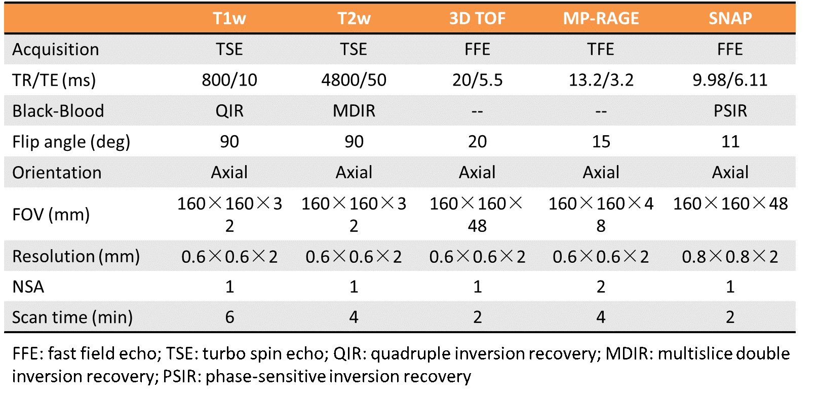
Study sample: Eighteen patients (mean age: 64.1 ± 6.1 years, 15 males) with carotid atherosclerotic disease (50%-70% symptomatic stenosis or >70% stenosis) referred to CEA were recruited. All patients underwent MR imaging within 1 week before surgery. MR imaging: Carotid MR imaging was performed on a 3.0T MR scanner (Achieva TX, Philips Healthcare, Best, The Netherlands) with 8-channel carotid coil. The MR imaging protocol included TOF, T1w, T2-weighted (T2w), MP-RAGE, and SNAP sequences. The imaging parameters are detailed in Figure 1. Histologic Sample Processing and Review: After CEA, the samples were sectioned (10μm) every 0.5mm throughout the length of the specimen and stained (hematoxylin and eosin [H&E]). The histologic specimens were reviewed and matched to the MR images by an experienced histologist and a radiologist who were blinded to MR image review with consensus. Morphological features of lumen, vessel wall, and calcifications were used for co-registration. The presence or absence and size of the IPHs were evaluated using ImageJ software. Image review: Two experienced radiologists blinded to histologic images interpreted the MR images which were matched to the histologic specimens with consensus by using Radiant software. The image review was conducted with two steps: 1) the reviewers outlined IPH on SNAP images blinded to MR-RAGE images; and 2) reviewers outlined IPH on MP-RAGE images blinded to SNAP images after one-month time interval to minimize memory bias. The IPH on MP-RAGE and SNAP images was defined as hyperintense compared to muscle (signal intensity ratio=1.5:1)4. Statistical analysis: The mean value of the number and the area of IPH measured by SNAP and MP-RAGE images and histology were calculated and compared. Shrinkage artifact induced by histological preparation was determined to be 7.8%5. The agreement of SNAP and MP-RAGE with histology in identifying IPH was determined using Cohen’s Kappa analysis. The agreement and associations of IPH area measured by MP-RAGE and SNAP images with histology were determined using Intra-class correlation coefficient (ICC), Pearson correlation coefficients (γ) and Bland-Altman analyses. The study protocol was approved by institutional review board and consent form was obtained from all subjects.Results
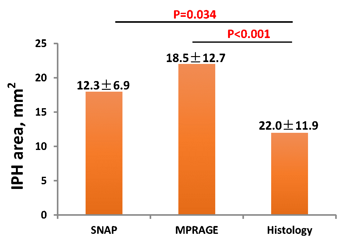
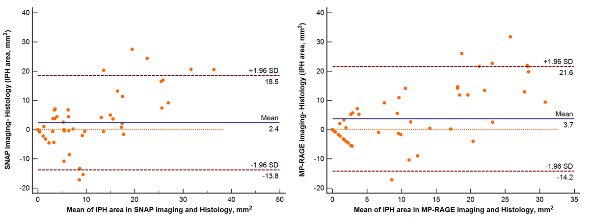
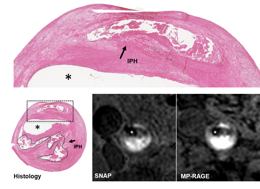
Of all 18 patients, 67 slices with acceptable image quality were eligible for MR and histologic image review. Of 67 slices, 44 (65.7%), 38 (56.7%), and 35 (52.2%) were found to have IPH on histology, SNAP, and MP-RAGE imaging, respectively (Fig 2). In identification of IPH, the Kappa value for SNAP and MP-RAGE images (agreement with histology) was 0.69 (standard error, 0.032) and 0.43 (standard error, 0.109) (Fig 2). For all slices (n=29) with IPH detected by SNAP and MP-RAGE images and histology, the area of IPH measured by SNAP (18.5±12.7 mm2, p=0.034) and MP-RAGE (22.0±11.9 mm2, p<0.001) (Fig. 3). Good agreement was found in measuring the area of IPH between SNAP images and histology (ICC, 0.795; 95% CI, 0.667-0.874) and between MP-RAGE and histology (ICC, 0.747; 95% CI, 0.554-0.833). Bland-Altman analysis revealed that, in measuring the area of IPH, the bias of SNAP (mean value, 2.4mm2) was smaller than that of MP-RAGE (mean value, 3.7mm2) compared to histology (Fig. 4). Figure 5 represents examples showing IPH on MP-RAGE and SNAP images and histologic section.Discussion and Conclusion
In this study, we investigated the performance of SNAP and MPRAGE imaging in identifying carotid IPH validated by histology. In identification and quantification of carotid IPH, SNAP showed better agreement with histology compared with MP-RAGE imaging. This might be due to the larger dynamic range and higher IPH-wall contrast-to-noise ratio of SNAP compared with MP-RAGE6.Our results suggest that SNAP imaging might be a more sensitive and accurate imaging approach in assessing carotid IPH than MP-RAGE imaging.Acknowledgements
None.References
1. Saam T, Hetterich H, Hoffmann V, et al. Meta-analysis and systematic review of the predictive value of carotid plaque hemorrhage on cerebrovascular events by magnetic resonance imaging. J Am Coll Cardiol. 2013;62:1081-1091.
2. Yamada K, Yoshimura S, Kawasaki M, et al. Embolic complications after carotid artery stenting or carotid endarterectomy are associated with tissue characteristics of carotid plaques evaluated by magnetic resonance imaging. Atherosclerosis. 2011;215:399-404.
3. Ota H, Yarnykh VL, Ferguson MS, et al. Carotid intraplaque hemorrhage imaging at 3.0-T MR imaging: comparison of the diagnostic performance of three T1-weighted sequences. Radiology. 2010;254:551-563.
4. Li D, Zhao H, Chen X, et al. Identification of intraplaque haemorrhage in carotid artery by simultaneous non-contrast angiography and intraplaque haemorrhage (SNAP) imaging: A magnetic resonance vessel wall imaging study. Eur Radiol. 2017 Nov 2. [Epub ahead of print]
5. Gamble G, Beaumont B, Smith H et al. B-mode ultrasound images of the carotid artery wall: correlation of ultrasound with histological measurements. Atherosclerosis 1993;102:163-173
6. Wang J, Börnert P, Zhao H, et al. Simultaneous noncontrast angiography and intraplaque hemorrhage (SNAP) imaging for carotid atherosclerotic disease evaluation. Magn Reson Med. 2013;69:337-345.
Figures