2080
Plaque permeability assessed with dynamic contrast-enhanced MRI predicts ferumoxytol nanoparticle delivery in patients with peripheral artery disease1Department of Vascular Medicine, Amsterdam UMC, Amsterdam, Netherlands, 2Department of Biomedical Engineering and Physics, Amsterdam UMC, Amsterdam, Netherlands, 3TMII, Icahn School of Medicine, New York, NY, United States, 4University Hospital Erlangen, Erlangen, Germany, 5Department of Radiology and Nuclear Medicine, Amsterdam UMC, Amsterdam, Netherlands
Synopsis
We investigated whether dynamic-contrast enhanced (DCE-)MRI could be used to predict accumulation of ferumoxytol nanoparticles in femoral plaques. To this end, we implemented USPIO enhanced quantitative T2* imaging, as well as 3D black-blood DCE-MRI sequences for femoral artery vessel wall imaging. Patients with peripheral artery disease (PAD) and healthy volunteers were included in this study. We show that ferumoxytol nanoparticle delivery to atherosclerotic plaques is associated with plaque permeability as assessed with DCE-MRI in patients with PAD.
Introduction
Nanomedicine has offered the promise of improving drug delivery to target organs, minimizing systemic adverse effects while maximizing local efficacy.(1) Because adequate delivery of drug-loaded nanoparticles to the atherosclerotic plaque is required to achieve a therapeutic response, and such therapy is likely to be costly, identifying which patients (or plaques) are amenable to nanotherapy is essential. Preclinical models have shown that nanoparticle translocation across the endothelium is dependent on microvascular permeability.(2)
Ultrasmall superparamagnetic iron-oxide particles (USPIO; ferumoxytol) are long circulating nanoparticles which are taken up by tissue macrophages.(3) Accumulation of particles can be assessed using MRI by quantifying its effect on the local R2* relaxation values. In this study, we hypothesized that accumulation of ferumoxytol nanoparticles in plaques could be predicted by assessing plaque permeability using dynamic-contrast enhanced (DCE-)MRI. To this end, we implemented a novel 3D black-blood DCE-MRI technique with high spatiotemporal resolution, allowing for accurate quantification of vessel wall contrast enhancement. We combined both imaging modalities in patients with peripheral artery disease (PAD) to assess whether USPIO uptake in femoral plaques was related to plaque permeability.
Methods
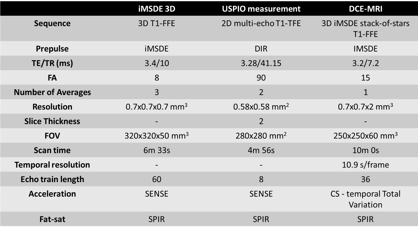
Approval for this study was obtained from our local medical ethical committee. We included 18 patients with PAD, as well as 8 healthy controls. MRI was performed on a 3T system (Philips Ingenia, Best, the Netherlands). Detailed scan parameters for all sequences are summarized in Figure 5. High-resolution black-blood anatomical images were made using a 3D IMSDE prepared black-blood gradient echo. To assess USPIO uptake, T2* mapping was performed in slices with visible plaque using a multi-echo gradient echo sequence, before (baseline) and 72 hours post- USPIO injection (ferumoxytol 4 mg/kg; AMAG Pharmaceuticals). R2* maps were calculated from the multi-echo data, and manually segmented in plaque, vessel wall, and lumen regions. For 3D black-blood DCE-MRI, we used a golden-angle stack-of-stars sequence, with an iMSDE black-blood pre-pulse, and a compressed sensing reconstruction into timeframes of 11 sec/frame, which was presented previously.(4) DCE-MRI was performed continuously during which volunteers were injected with contrast agent (Gadovist, 0.1 mmol/kg) four minutes into the scan. Area under the curve (AUC) values were calculated from the dynamic contrast images, covering the two minutes following contrast injection.
Results & Discussion
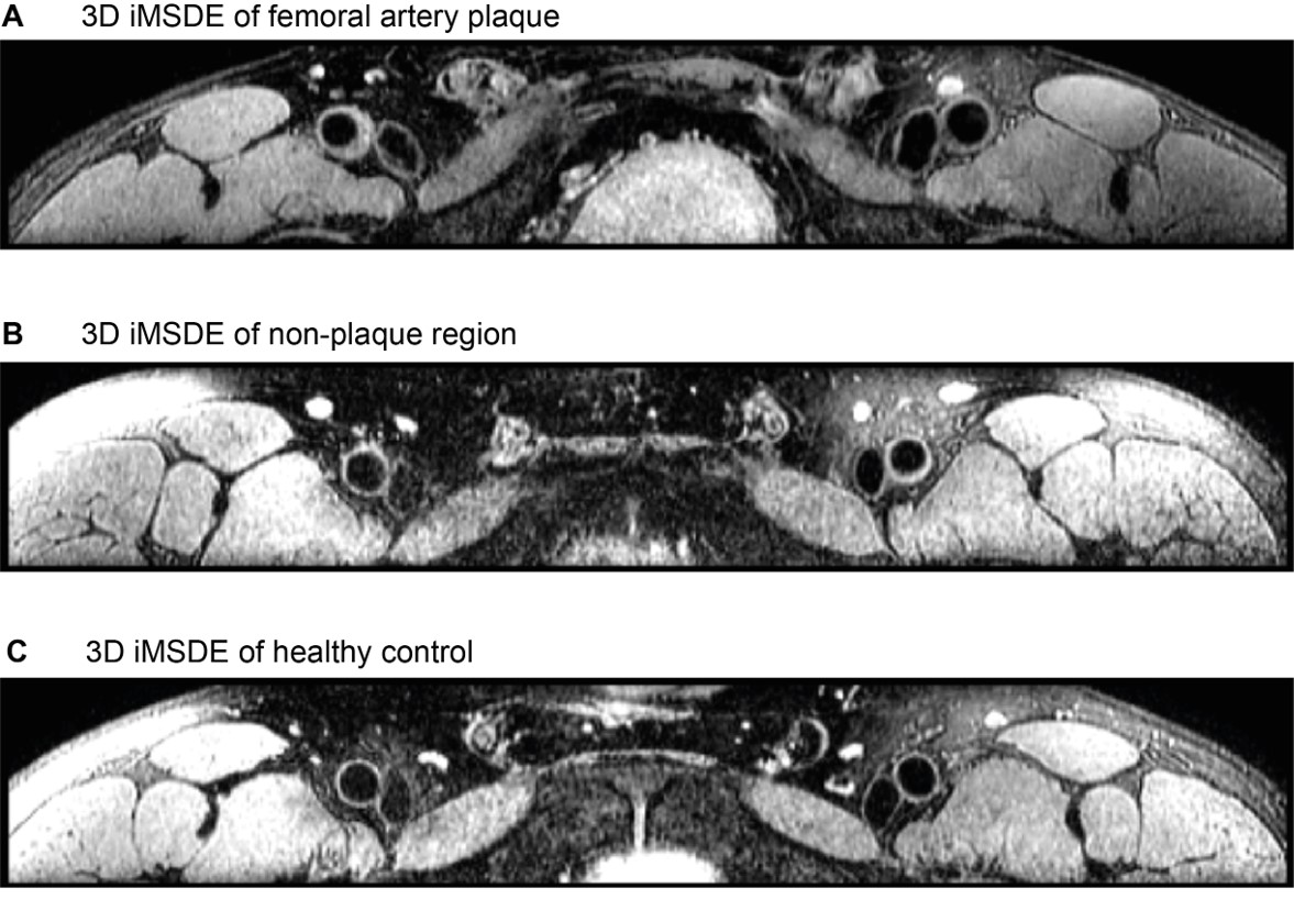
Figure 1 shows representative images of identified plaque (A) and non-plaque segment (B) of the femoral arteries in a patient, as well as in a healthy control (C). Despite the challenging anatomy, SNR and blood suppression was very good along most of the length of the femoral artery. Vessel wall thickness of plaques was significantly higher (1.90 mm) as compared to non-plaque regions in patients (1.15 mm; p<0.001) and healthy controls (0.94 mm; p<0.001).
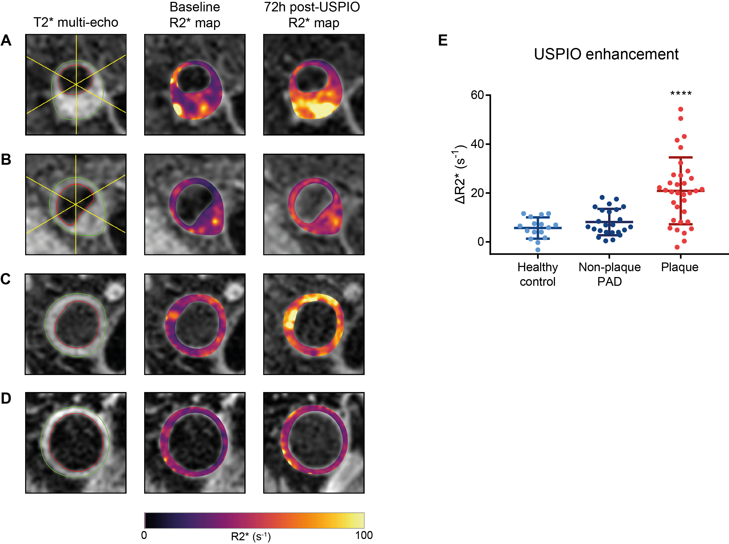
At baseline, R2* values of plaque-containing segments were not different compared with non-plaque vessel wall in patients with PAD (44.3 ± 10.1 s-1 vs. 40.4 ± 5.8 s-1, p=0.153), but were slightly increased compared with the vessel wall of controls (35.5 ± 4.3 s-1, p=0.001). R2* enhancement of the vessel wall was detected by clear regional decreases in the R2* maps, especially in PAD patients (Figure 2A-C). Importantly, a wide spectrum of USPIO uptake patterns could be detected: Figure 2A depicts strong enhancement of plaque; Figure 2B shows low enhancement of plaque; Figure 2C demonstrates moderate enhancement of a non-plaque segment. R2* changes in plaque segments (ΔR2* = 19.9±12.5 s-1) was markedly increased compared with non-plaque areas (ΔR2* = 8.1±5.4 s-1; p<0.001) and controls (ΔR2* = 5.67 ± 4.38 s-1; p<0.001; Figure 2D). (Figure 2E)
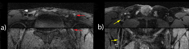
DCE-MRI also revealed a wide range of contrast enhancement patterns, with some plaques showing clear enhancement, while others showing little change in image intensity over time (Figure 3). Importantly, good blood suppression was maintained throughout, enabling accurate delineation of plaque, vessel wall and lumen.
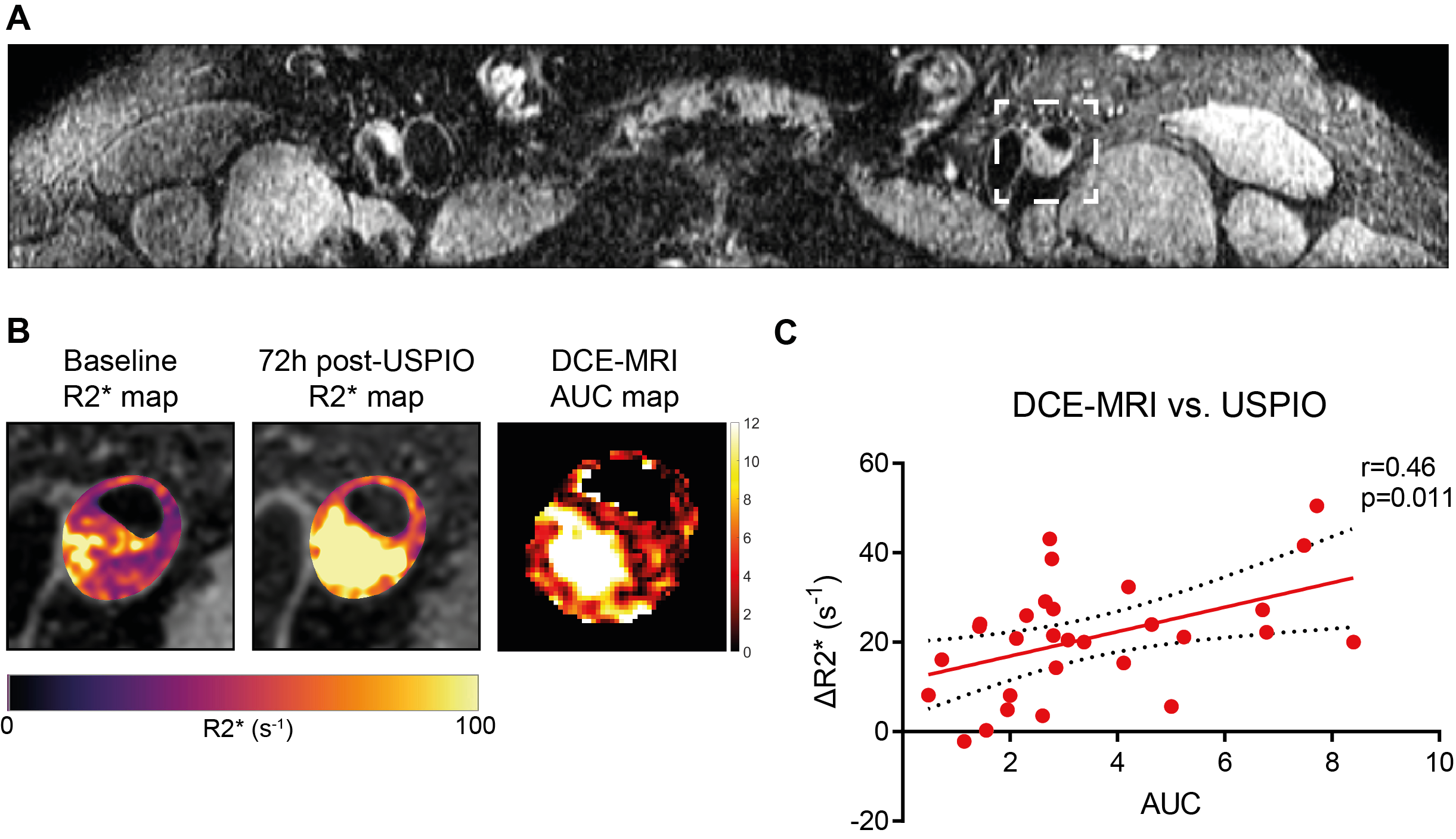
In patients with PAD, AUC maps showed co-localization with USPIO uptake as indicated by the R2* maps of the corresponding plaques (Figure 4A-B). More importantly, for plaque segments, there was a significant correlation between the AUC and ΔR2* (r=0.52; p=0.002) (Figure 4C). In a multivariable analysis, adjusting for age, sex, BMI, smoking, diabetes, hypertension, LDL-c and Lp(a), the AUC remained a significant predictor of ΔR2* (ß 3.24 (1.00-5.49), p=0.007).
Conclusion
We demonstrate that MRI detected ferumoxytol nanoparticle delivery to atherosclerotic plaques is associated with plaque permeability as assessed with DCE-MRI in patients with PAD. We envision that further development of these imaging modalities may assist in evaluation of novel therapeutics and monitoring of disease progression in atherosclerosis.
Acknowledgements
We thank all patients and healthy volunteers for their participation.References
1. Lobatto ME, Fuster V, Fayad ZA, Mulder WJ. Perspectives and opportunities for nanomedicine in the management of atherosclerosis. Nat Rev Drug Discov 2011;10:835-52.
2. Kim Y, Lobatto ME, Kawahara T et al. Probing nanoparticle translocation across the permeable endothelium in experimental atherosclerosis. Proc Natl Acad Sci U S A 2014;111:1078-83.
3. Sadat U, Usman A, Gillard JH. Imaging pathobiology of carotid atherosclerosis with ultrasmall superparamagnetic particles of iron oxide: an update. Curr Opin Cardiol 2017;32:437-440.
4. Schoormans J, Zheng KH, Stroes ES, Strijkers G, Nederveen AJ, Coolen BF. 3D black-blood DCE-MRI using radial stack-of-stars acquisition and CS reconstruction: application in carotid and femoral arteries. Proceedings of the 25th Annual Meeting of the ISMRM, Honolulu, 2017.
Figures