2079
A lightweight and ultra-flexible “blanket” coil design for carotid artery wall imaging1Weill Cornell Medicine, New York, NY, United States, 2New York University, New York, NY, United States
Synopsis
We aimed to build an ultra-flexible blanket coil for carotid wall imaging. Results in healthy volunteers showed that the coil improves comfort and allows easy positioning in the neck while providing 32% higher SNR at the carotid bifurcation compared to the product 64-channel neurovascular coil.
INTRODUCTION
MRI plays an important role in the assessment of carotid plaque components for predicting future stroke risk (1,2). Carotid plaque and vessel wall MRI requires submillimeter resolution within short scan time to minimize motion artifacts caused by bulk motion, coughing, or swallowing. An effective approach is to use multi-channel surface coils designed to maximize SNR at the carotid bifurcation where most plaque buildups occur (3). However, these dedicated carotid coils are often bulky and provide limited coverage which makes coil positioning more difficult when compared to the standard neurovascular coil (4). Our objective was to design an ultra-flexible coil that allows easy customized positioning and improves patient comfort while providing SNR benefit over the neurovascular coil.METHODS
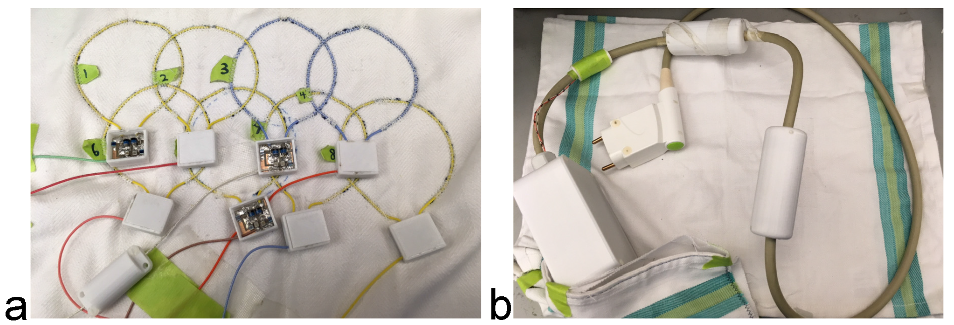
Blanket coil design. We built a lightweight and ultra-flexible “blanket” receive array out of eight high-impedance coil elements using micro-coaxial cables (Fig.1) (5). The coils were sewed to a thin fabric substrate such that the ensemble device allowed a tailored fit on potentially cumbersome anatomy such as the carotid arteries. Slim 3D printed housing units protected the interface boards, which each contained two pin diodes (MA4P4002B-402; Macom, Lowell, MA, USA) to detune the receive coils during body coil excitation and a matching circuit whose output was connected to a coaxial cable with the proper length for “reversed” preamplifier decoupling (5). The cables included inline traps to suppress shield currents and a fuse was implemented in each coil for patient safety.
Carotid MRI experiment. Five healthy volunteers were scanned with the blanket coil and also with the commercially available 64-channel neurovascular (NV64) coil on a Siemens Prisma 3T MRI system. The imaging protocol consisted of 2D black blood T1w turbo spin echo sequence (0.63x0.63x2 mm3 voxel size, 885 ms TR, 9.4 ms TE) for carotid wall visualization and 3D multi-echo GRE sequence (0.6x0.6x3 mm3 voxel size, 21 ms TR, 4.7 ms echo spacing, 10° flip angle) for quantitative susceptibility mapping (QSM). The imaging volume was approximately 6 cm thick centered on the carotid bifurcation. Map in absolute SNR unit for each coil was computed from two single-slice 2D GRE scans of the carotid bifurcation with TR, TE, and flip angle matching those of 3D QSM sequence and RF excitation turned on (for signal measurement) and off (for noise reference measurement) following the procedure outlined in (6).
RESULTS
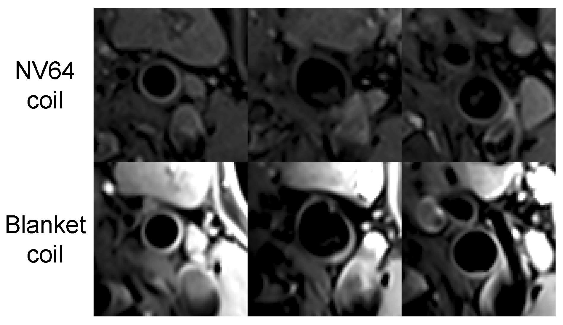
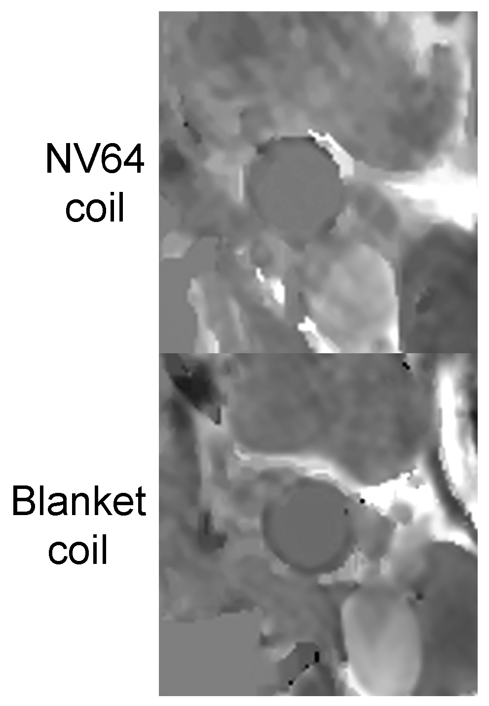
Figure 2 shows an example of black blood T1w images obtained with the product NV64 and proposed blanket coils, demonstrating consistently improved visualization of the carotid artery wall before, at, and after the carotid bifurcation. Similarly, QSM depiction of the carotid wall was improved with the blanket coil (Fig.2). Overall, the SNR of the blanket coil measured at the level of the carotid bifurcation was 32% higher than that of NV64 coil (29.7 ± 6.7 vs. 22.5 ± 6.1).DISCUSSION
Our preliminary results showed that the proposed lightweight and ultra-flexible blanket coil were well tolerated by the volunteers and provided easy positioning as well as better SNR than the neurovascular coil for carotid wall imaging. The proposed design can be translated to other body parts with irregular shapes and further evaluation in patients in the clinical setting is warranted.Acknowledgements
No acknowledgement found.References
1. Cai JM, Hatsukami TS, Ferguson MS, Small R, Polissar NL, Yuan C. Classification of human carotid atherosclerotic lesions with in vivo multicontrast magnetic resonance imaging. Circulation 2002;106(11):1368-73.
2. Takaya N, Yuan C, Chu B, Saam T, Underhill H, Cai J, Tran N, Polissar NL, Isaac C, Ferguson MS, Garden GA, Cramer SC, Maravilla KR, Hashimoto B, Hatsukami TS. Association between carotid plaque characteristics and subsequent ischemic cerebrovascular events: a prospective assessment with MRI--initial results. Stroke 2006;37(3):818-23.
3. Balu N, Yarnykh VL, Scholnick J, Chu B, Yuan C, Hayes C. Improvements in carotid plaque imaging using a new eight-element phased array coil at 3T. J Magn Reson Imaging 2009;30(5):1209-14.
4. Brinjikji W, DeMarco JK, Shih R, Lanzino G, Rabinstein AA, Hilditch CA, Nicholson PJ, Huston J 3rd. Diagnostic accuracy of a clinical carotid plaque MR protocol using a neurovascular coil compared to a surface coil protocol. J Magn Reson Imaging 2018;48(5):1264-1272.
5. Zhang B, Sodickson DK, Cloos MA. A high-impedance detector-array glove for magnetic resonance imaging of the hand. Nature Biomedical Engineering 2018;2(8):570-577.
6. Kellman P, McVeigh ER. Image reconstruction in SNR units: a general method for SNR measurement. Magn Reson Med 2005;54(6):1439-1447.
Figures