2073
Whole heart coronary MRA using non-selective balanced SSFP sequence at 3.0T: comparison of image qualityIsao Shiina1, Michinobu Nagao2, Masami Yoneyama3, Kazuo Kodaira1, Yasuhiro Goto1, Yoshihiro Ikeda1, Yutaka Hamatani1, Mamoru Takeyama1, Isao Tanaka1, and Shuji Sakai2
1Department of Radiological Services, Women's Medical University Hospital, Tokyo, Japan, 2Department of Diagnostic Imaging and Nuclear Medicine, Women's Medical University Hospital, Tokyo, Japan, 3Philips Electronics Japan, Tokyo, Japan
Synopsis
3 tesla derived MRCA has a limitation of low contrast for coronary arteries because of setting lower flip angles due to high SAR. Therefore, the signal contrast of for coronary arteries becomes low. 3D non-selective balanced TFE (3D nsbTFE) has the possibility to solve this problem. We examine image quality of MRCA obtained from 3 tesla and 3D nsbTFE.
Synopsis
3 tesla derived MRCA has a limitation of low contrast for coronary arteries because of setting lower flip angles due to high SAR. Therefore, the signal contrast of for coronary arteries becomes low. 3D non-selective balanced TFE (3D nsbTFE) has the possibility to solve this problem. We examine image quality of MRCA obtained from 3 tesla and 3D nsbTFE.INTRODUCTION
Coronary MR angiography (MRCA) is widely used for the assessment of coronary artery disease, and has big merits of no radiation exposure and unnecessary of contrast media. (REF.1)From the view point of magnetic field strength, 1.5 tesla scanner for coronary MRA is more used in clinical than 3 tesla scanner because MRCA from balanced sequence with 1.5 tesla has a good contrast in signal intensity between blood and myocardium. On the other hand, 3 tesla has to set up lower flip angles for MRCA due to a limitation of high SAR. Therefore, the signal contrast of for coronary arteries becomes low (REF.2) if T1TFE sequence is used. Furthermore, MRCA at 3 tesla has a serious problem of the banding artifacts due to B0 inhomogeneity (REF.3). 3D non-selective balanced TFE (3D nsbTFE) has the possibility to solve this problem. 3D nsbTFE enables shorter TR by not using the slice selective excitation (Fig.1), leading to setting up high flip angles. Therefore, we hypothesized that 3D nsbTFE can obtain high contrast for coronary arteries without artifact and less local shimming.Methods
Using 3.0T MR system (Ingenia, Philips Electronics Japan) and torso-cardiac coil, data of MRCA for 5 healthy volunteers was analyzed. We compared image quality among three sequences of the conventional method (TFE), the balanced SSFP combined with T1 TFE (b-TFE), and 3D nsbTFE. Three sequences were acquired with navigator and cardiac triggering using the following parameters; TFE, FOV (mm) = 180×180, slice thickness (mm) = 1.5×1.5×1.5, TR/TE (ms) =3.4/1.51, flip angle (°) = 12, TFE factor = 29, NSA = 1, SENSE factor = 1×1, Slice = 160, Slice orientation = transverse, 100% gathering time (min) = 5:38; b-TFE, FOV (mm) = 180×180, slice thickness (mm) = 1.5×1.5×1.5, TR/TE (ms) = 4.5/2.2, flip angle (°) = 70, TFE factor = 22, NSA = 1, SENSE factor = 1×1, Slice = 160, Slice orientation = transverse, 100% gathering time (min) = 8:11; nsbTFE, FOV (mm) = 280×280, slice thickness (mm) = 1.5×1.5×1.5, TR/TE (ms) = 2.3/0.93, flip angle (°) = 70, TFE factor=41, NSA=1, C-SENSE factor=7, Slice=330, Slice orientation = coronal,100% gathering time (min) = 3:04 (Fig.2). Curved Planer Reconstructions (CPRs) of MRCA were reconstructed using available software (Ziostation 2, Ziosoft Co, Tokyo) and image quality was evaluated by visual score at 10 places (RCA: #1,2,3,4 LAD: #5,6,7,8 CX: #11,13) based on American Heart Association classification (Fig. 3). The visual evaluation items were three items of overall image quality, sharpness, noise and artifacts, we evaluated them as 4-point grades (grade “4” was excellent, “1” was severe) by two blinded readers (one radiologist and one radiological technologist).Results & Discussion
Overall image quality and sharpness for 3D nsbTFE were significantly higher than those for TFE and bTFE. On the other hand, the noise artifact for 3D nsbTFE was significantly decreased than that for TFE and bTFE. The signal noise ratio for 3D nsbTFE was higher than TFE and bTFE. The banding artifacts were often seen on bTFE. Three branches of coronary arteries could not be detected on bTFE. 3D nsbTFE successes to shorten and can set up high flip angle. Consequently, contrast for coronary arteries improved.Conclusion
MRCA using nsbTFE can overcome weak points in conventional balanced SSFP with 3 tesla while while ensuring the image quality and high signal to noise ratio.Acknowledgements
No acknowledgements found.References
(1) Danias PG,Roussakis A,loannidis JP,et al: Diagnostic performance of coronary magnetic resonance angiography as compared against conventional x-ray angiography: A meta-analysis.J Am Coll Cardiol. 2004 Nov 2; 44(9):1867-76. (2) Kaul MG, Stork A, Bansmann PM,et al: Evaluation of balanced steady-state free precession (TrueFISP) and K-space segmented gradient echo sequences for 3D coronary MR angiography with navigator gating at 3 Tesla.Rofo.2004 Nov:176(11):1560-5. (3)Thomas D1, Krug B, Hackmann D,et al: MR-coronary angiography: comparison of SSFP and spoiled GRE sequence (bright blood technique) and a TSE sequence (black blood technique) in healthy volunteers. Rofo. 2004 Nov:176(11):1589-98. (4) Ohyama K, Kubo H, Harada M,et al: Comparison of 3 Tesla whole heart coronary MRA (WHCA) with 1.5 Tesla. Nihon Hoshasen Gijutsu Gakkai Zasshi. 2008 Dec 20:64(12):1540-6.Figures
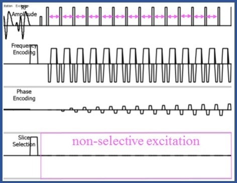
Figure 1: Sequence chart of non-selective TFE
With slice excitation as the whole, setting
of extremely short TR is enabled once by not performing it subsequently.
We
photograph non-selective TFE by Coronal collection of in consideration of an
artifact in reply to activate all in the boa to completely take a body in the
slice direction。
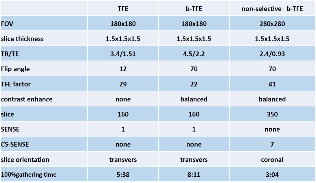
Table 1: Magnetic
resonance imaging sequences and parameters
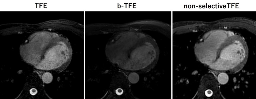
Figure 2: The thing which used
balanced together with a traditional approach in it, Ax image of non-selective
TFE. We treat it in Ax in non-selective TFE of the coronal collection of and
display it.
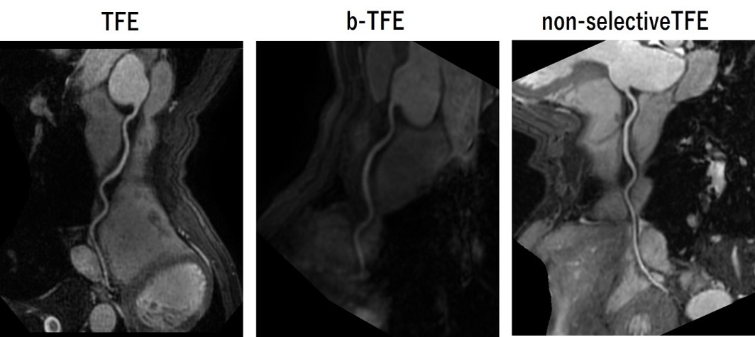
Figure 3: The
thing which used balanced together with a traditional approach in it, a CPR
image of non-selective TFE. High s/n in non-selective TFE stands out.
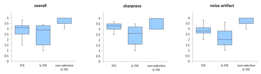
Figure 4: Results of overall, sharpness, noise artifacts in 5 volunteers