2055
Contrast-enhanced compressed sensing whole-heart 3T MR angiography in detection of coronary artery stenosis: A preliminary comparative study with computed tomography angiography1Peking Union Medical College Hospital, Peking Union Medical College, Chinese Academy of Medical Science, Beijing, China, 2Siemens Healthcare, MR Collaborations NE Asia, Beijing, China, 3Siemens Healthcare GmbH, Erlangen, Germany, Erlangen, Germany
Synopsis
The aim of this study was to investigate the diagnostic performance of contrast-enhanced compressed sensing (CS) whole-heart 3T MR angiography in the detection of clinical significant coronary artery stenosis by using CTA as a reference. The preliminary results indicate that the contrast-enhanced CS coronary MR angiography has a good consistency in evaluating coronary artery disease in comparison to CTA and a short enough acquisition time that can be applied in the waiting time between contrast injection and late gadolinium enhancement imaging.
INTRODUCTION
Coronary magnetic resonance angiography (MRA) is a promising noninvasive method for detecting coronary artery disease (CAD) which is radiation-free and not affected by severe calcification artifacts. However, the main limitation of the coronary MRA for clinical application is long acquisition time and limited resolution. Compressed sensing (CS) with sparse sampling and iterative reconstruction can effectively reduce acquisition time. Short acquisition time allows to achieve higher resolution due to high acceleration. The aim of this study was to evaluate the diagnostic performance of contrast-enhanced CS coronary MRA in the detection of clinically significant coronary artery stenosis by using CTA as a reference.METHODS
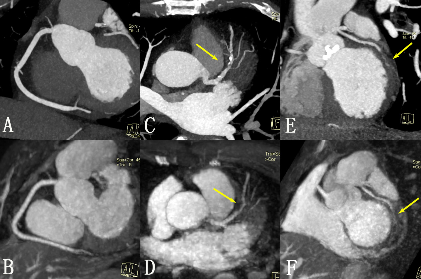
18 consecutive patients with clinically suspected CAD underwent contrast-enhanced CS coronary MRA followed by a CTA scan. All contrast-enhanced CS coronary MRA data were acquired on a 3T MR scanner (MAGNETOM Skyra, Siemens Healthcare, Erlangen, Germany). The key parameters of the T2-prepared GRE protoype sequence were as follows: TR/TE = 4/1.7ms, T2 prep.duration=50ms, FA=20deg, bandwidth=401Hz/Px, voxel size=1.1 x 1.1 x 1.1 mm3, acceleration factor 10.21. The quality of the contrast-enhanced CS MRA image was graded for each segment on a four-point scale (1: poor, 2: fair, 3: good; 4: excellent). All the segments with quality scores 2–4 were assessed to identify significant narrowing (≥50% lumen diameter reduction) in comparison with CTA. The inter-modality agreement between MRA and CTA evaluation of coronary artery stenosis was assessed using a Kappa test. SPSS (version 20, IBM, America) was used for data analysis.RESULTS
All 18 patients underwent contrast-enhanced CS MRA successfully with the mean heart rate of 68±15 beats per minute. The average imaging time was 5.8±1.9 minutes. Among 162 segments in total, 146 (90.1%) segments had a diagnostic image quality and were included in the analysis. The inter-modality evaluation between MRA and CTA was 0.708 (p<0.01), representing good agreement for assessment of stenosis.DISCUSSION
In this study, CS whole-heart coronary MRA was obtained with a substantially reduced acquisition time (5.8±1.9min) compared to previous studies using conventional coronary MRA2 which indicating a high isotropic resolution achieved. Between contrast injection and late gadolinium enhancement imaging there is typically a 8-10min waiting time. This study demonstrated that our CS MRA acquisition could be applied successfully in the waiting time. The contrast-enhanced CS coronary MRA showed a good consistency in detecting CAD compared with CTA indicating an effective potential alternative to rule out significant coronary artery stenosis without exposure to ionizing radiation. The limitation of this study includes a small sample size and the lack of invasive coronary angiography as gold standard to accurately calculate the diagnostic performance. Further large-sample studies and image quality improvements are required.CONCLUSION
Contrast-enhanced CS whole-heart coronary 3T MR angiography is a promising noninvasive technique for evaluating clinically significant coronary stenosis with a shorter acquisition time.Acknowledgements
No acknowledgement found.References
1. Liu J. et al.; Dynamic cardiac MRI reconstruction with weighted redundant Haar wavelets; Proc. ISMRM 2012, #178.
2. Yang Q, Li K, Liu X, et al. 3.0T whole-heart coronary magnetic resonance angiography performed with 32-channel cardiac coils: a single-center experience. Circulation Cardiovascular imaging. 2012;5(5):573-579
Figures