2045
Characterization of tissue properties of tissue engineered cardiac patches using DTI at 22.3T1Department of Radiology, University Medical Center Utrecht, Utrecht, Netherlands, 2Department of Cardiology, Experimental Cardiology, University Medical Center Utrecht, Utrecht, Netherlands, 3Department of Orthopaedics, University Medical Centre, Utrecht, Netherlands, 4Department of Biomedical Engineering, Eindhoven University of Technology, Eindhoven, Netherlands, 5Department of Equine Sciences Faculty of Veterinary Sciences, Utrecht University, Utrecht, Netherlands
Synopsis
Engineering three-dimensional (3D) tissues with similar properties to native myocardium offers a promising approach to restore cardiac function after myocardial infarction. However, visualizing the orientation of the tissue in tissue engineered cardiac patches using immunofluorescent imaging has proven difficult due to the 3D and dense tissue structure. In this study, we have applied diffusion tensor imaging at 22.3T at ultra-high resolution (62.5μm isotropic) to characterize the tissue properties of cardiac patches. We show that adding fibroblasts induces cellular organization of stem cell-derived cardiomyocytes in the patch, resulting in more diffusion restriction and higher anisotropy, better mimicking native myocardial tissue properties.
Introduction
True myocardial regeneration or repair can only be achieved by replacing the myocardial tissue that dies during a myocardial infarction. Engineering three-dimensional (3D) tissues with similar properties to the native myocardium offers a promising approach to restore cardiac function. To this end we have engineered cardiac patches using melt electrowriting (MEW) to recreate the fibrous structure of the native extracellular matrix and combined these scaffolds with human induced pluripotent stem cell-derived cardiomyocytes (iPSC-CM) with or without cardiac fibroblasts in a collagen-rich hydrogel (1). However, visualizing the orientation of the tissue using immunofluorescent imaging has proven difficult due to the 3D and dense tissue structure. In this study, we have applied diffusion tensor imaging (DTI) at 22.3T to characterize the tissue properties of tissue engineered cardiac patches.Methods
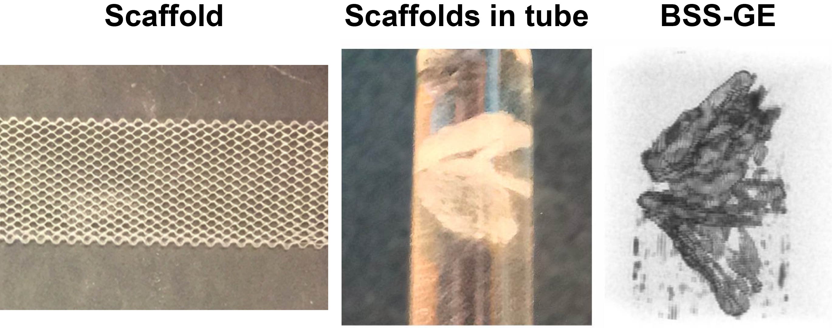
Using previously described methods, scaffolds with hexagonal microarchitectures were fabricated using melt electrowriting (1). Hexagonal MEW scaffolds with 400 μm side lengths and 350 μm thickness were seeded with either iPSC-CMs (scaffold 1 and 2), or iPSC-CMs and 10% human cardiac fibroblasts (FBs) (scaffold 3, 4 and 5) in a collagen/agarose-based hydrogel (30,000 cells per μl hydrogel). The engineered cardiac patches were then cultured in vitro for one week and subsequently fixed in 4% paraformaldehyde for 15 minutes. MR samples were prepared by punching out 4 mm diameter patches, which were submerged in perfluoropolyether (Galden, Solvay) in a 5 mm NMR tube.
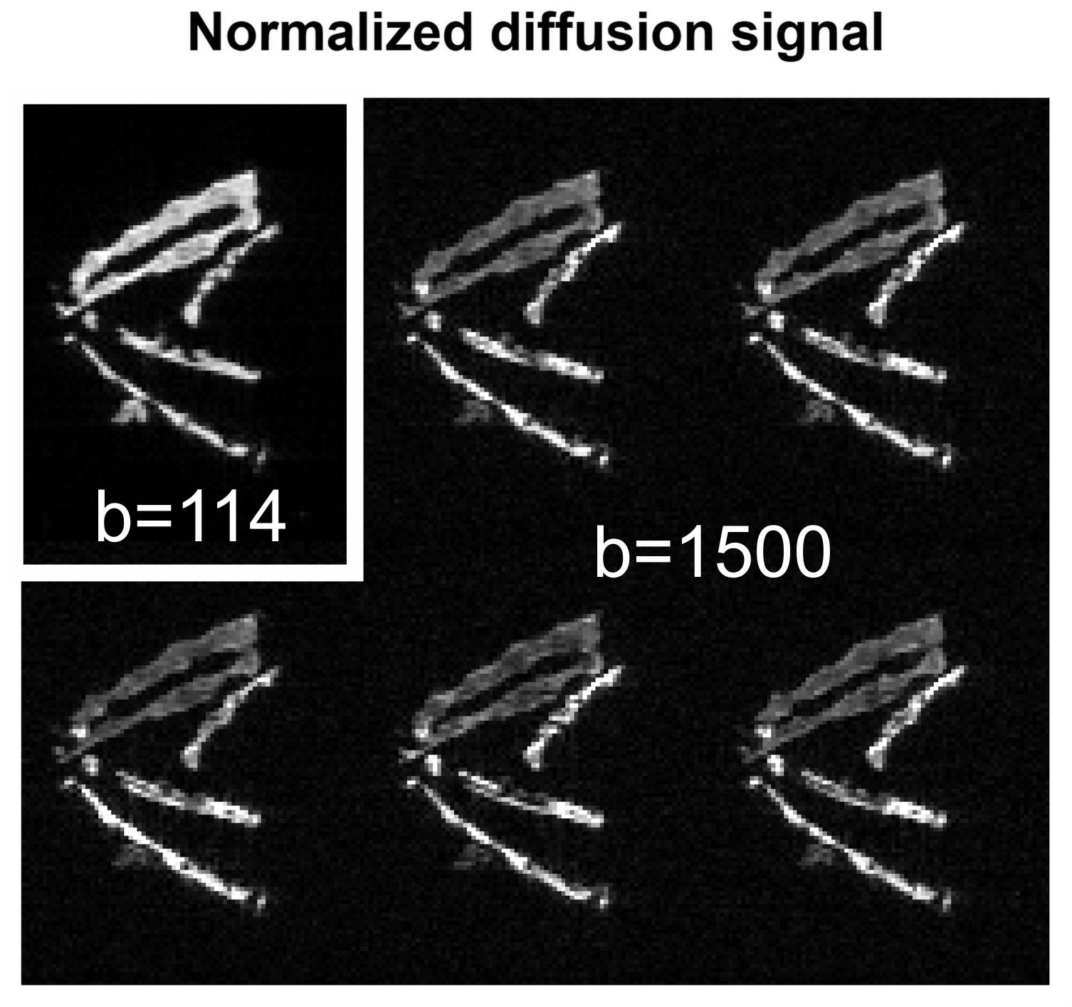
MRI acquisition was performed on a 22.3T (950 MHz) vertical bore system (Bruker) at room temperature (293 K). First a 3D balanced steady-state gradient echo (BSS-GR) acquisition with a resolution of 25x25x25 μm3 and a matrix size of 200x260x200 (TE = 2.35 ms; TR = 4.7 ms; NSA=64) was performed. Secondly, a diffusion weighted spin-echo (DW-SE) acquisition with a resolution of 62.5x62.5x62.5 μm3 and a matrix size of 80x104x80 (TE = 16 ms; TR = 1500 ms; NSA = 1; acq time = 2 h per diffusion direction) was performed. The diffusion measurement consisted of 3 volumes with b=114 s/mm2 and 24 volumes with b=~1500 s/mm2 and the entire measurement was performed twice.
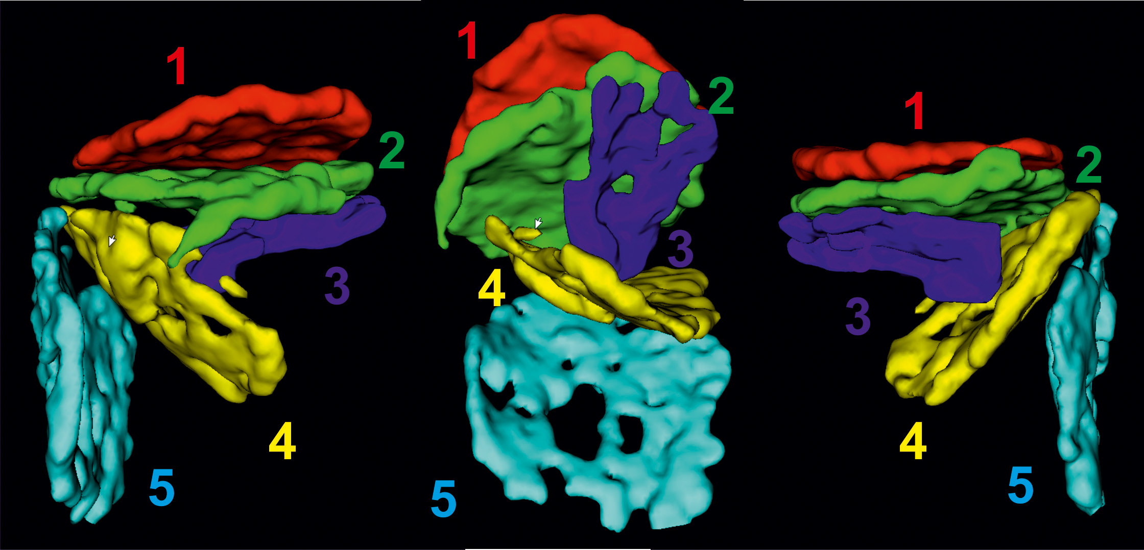
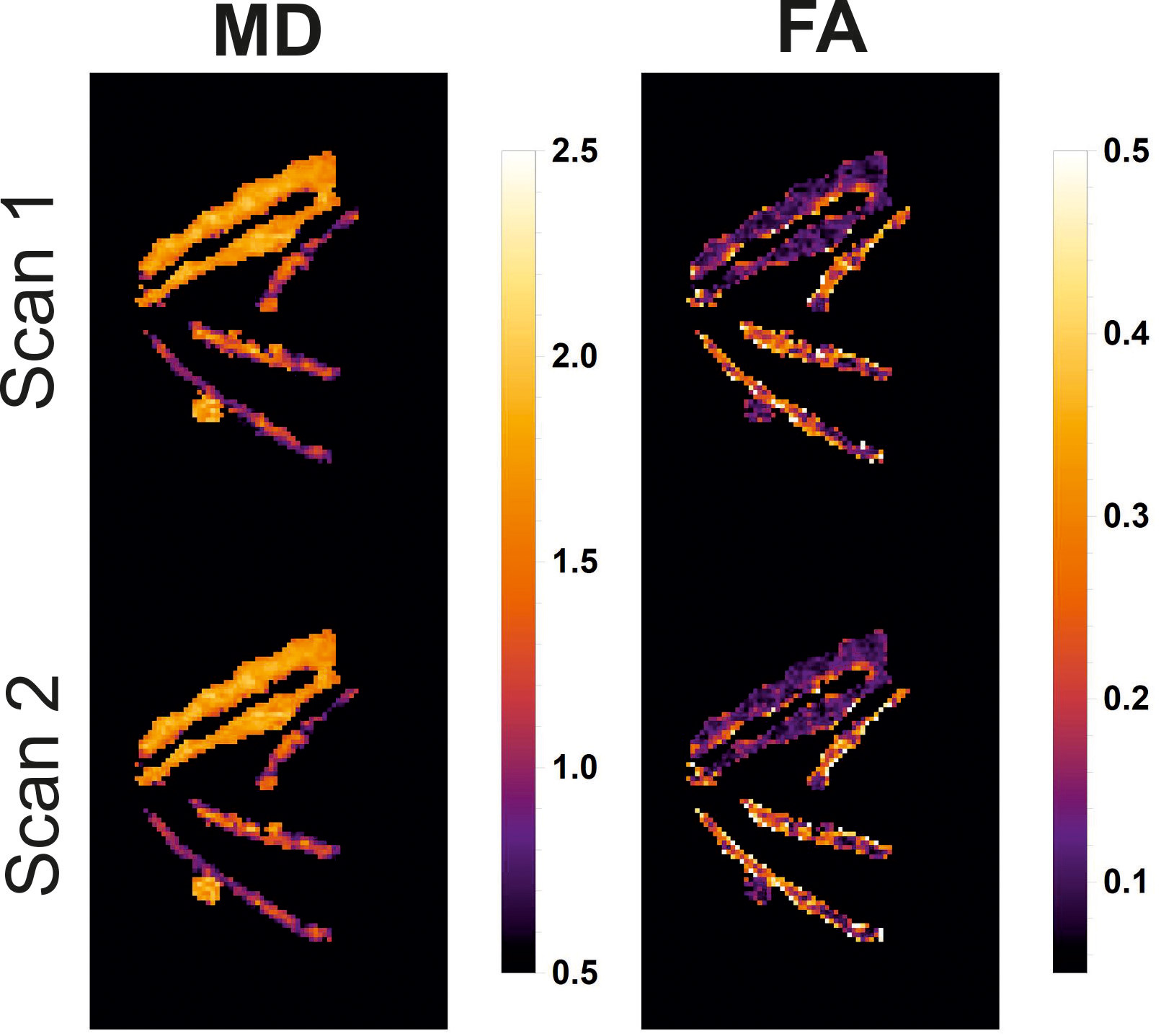
Data was processed using DTITools for Mathematica (github.com/mfroeling/DTITools). First the Bruker data was converted to the nifty file format using Bruker2NifTi (github.com/neurolabusc/Bru2Nii). The diffusion tensor was fitted using an iterative weighted linear least squares solver (iWLLS) (2) with outlier rejection using REKINDLE (3). From the diffusion tensor eigenvalues (λ1, λ2, λ3) the mean diffusivity (MD) and fractional anisotropy (FA) were calculated. The scaffolds were manually segmented using ITKSnap (itksnap.org). Additionally, a threshold mask of the mean DWI data was made. By multiplying both masks, it was assured that only parameters within each scaffold were measured. For each scaffold and dataset the mean values and standard deviation of the tensor eigenvalues, MD and FA were calculated.
Restults
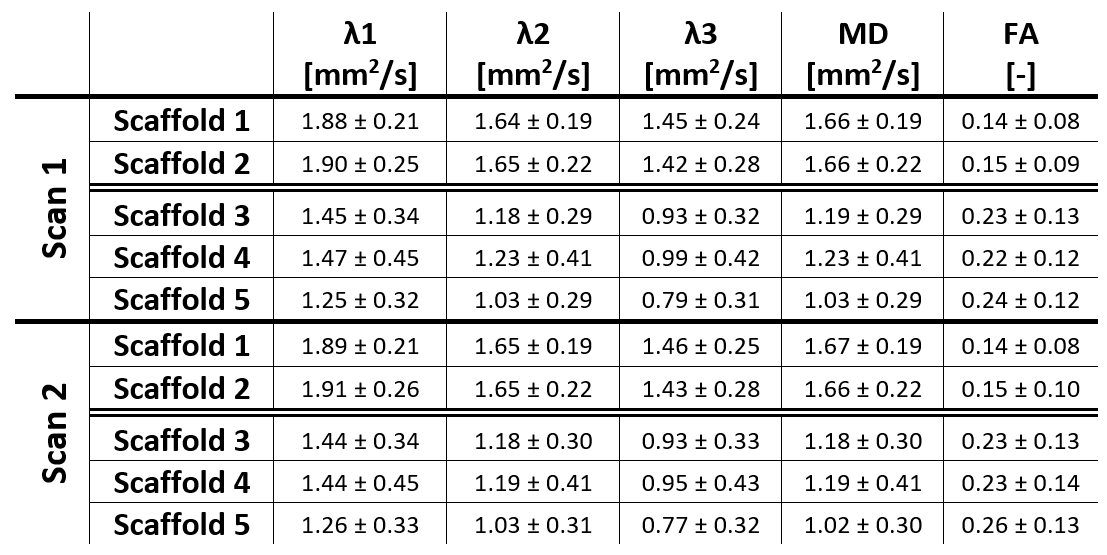
Fig. 1 shows the scaffold and a maximum intensity projection of the BSS-GE data and Fig. 2 shows cross sections of the DW-SE data for b=114 s/mm2 and b=1500 s/mm2. The segmentation of each scaffold is shown in Fig. 3. After tensor estimation, the diffusion parameters were calculated and quantitative maps of MD and FA are shown in Fig. 4. Both from the raw diffusion data as well as the MD and FA there is a clear distinction between scaffolds 1 and 2 compared to scaffolds 3, 4 and 5. Quantitative values for all diffusion parameters are reported in Table 1. Scaffolds 1 and 2, containing iPSC-CMs only, had an MD of ~1.66 mm2/s and an FA of ~1.5, indicating that the diffusion is hindered but no real anisotropy is observed. Scaffolds 3, 4 and 5, containing iPSC-CMs and FBs, had an MD of ~1.14 mm2/s and an FA of ~0.24 indicating more diffusion restriction and higher anisotropy. The values of the last three scaffolds closely resemble those of in-vivo and ex-vivo measurements of myocardial tissue (4).Conclusion
At the ultra-high resolution achievable at 22.3T, DTI proves to be a powerful method to characterize tissue properties of tissue engineered cardiac patches. We have shown that adding fibroblasts induces cellular organization of the iPSC-CMs, resulting in more diffusion restriction and higher anisotropy, better mimicking native myocardial tissue properties.Acknowledgements
Experiments at the 950 MHz instrument were supported by uNMR-NL, an NWO-funded National Roadmap Large-Scale Facility of the Netherlands (project 184.032.207).References
1. Castilho M and van Mil A, Maher M, Metz CHG, Hochleitner G, Groll J, Doevendans PA, Ito K, Sluijter JPG, Malda J: Melt electrowriting allows tailored microstructural and mechanical design of scaffolds to advance functional human myocardial tissue formation. Adv Funct Mater 2018, 1803151.
2. Veraart J, Sijbers J, Sunaert S, Leemans A, Jeurissen B: Weighted linear least squares estimation of diffusion MRI parameters: Strengths, limitations, and pitfalls. Neuroimage 2013; 81:335–346.
3. Tax CMW, Otte WM, Viergever MA, Dijkhuizen RM, Leemans A: REKINDLE: Robust Extraction of Kurtosis INDices with Linear Estimation. Magn Reson Med 2015; 73:794–808.
4. Rose JN, Nielles-Vallespin S, Ferreira PF, Firmin DN, Scott AD, Doorly DJ: Novel insights into in-vivo diffusion tensor cardiovascular magnetic resonance using computational modelling and a histology-based virtual microstructure. Magn Reson Med 2018.
Figures