2041
In Vivo Potassium MRI of the Human Heart at 7.0 Tesla1Max Delbrueck Center for Molecular Medicine, Berlin, Germany, 2Institute of Radiology, University Hospital Erlangen, Erlangen, Germany, 3Division of Medical Physics in Radiology, German Cancer Research Center (DKFZ), Heidelberg, Germany, 4Institute of Medical Physics, University of Erlangen, Erlangen, Germany, 5MRI.TOOLS GmbH, Berlin, Germany
Synopsis
Potassium ions (K+) play a critical role in cardiac electrophysiology; changes in their concentration might reflect ongoing pathophysiological processes related to cardiovascular diseases. In vivo potassium (39K) MRI, due to its extremely low sensitivity remains a major challenge. Here we investigated, for the first time, the feasibility of in vivo 39K MRI of human heart in a healthy subject. To achieve this goal, we developed a custom-built 39K/1H RF coil, which is tailored for 39K MRI of human heart at 7.0 Tesla. This approach facilitated 39K MRI of human heart with an isotropic spatial resolution of 14.5 mm3 within a total scan time of 30 minutes.
Introduction
Potassium channels represent the largest and most functionally-diverse family of ion-channel proteins in the human genome1. Sodium/potassium pumps (Na+/K+-ATPase) play a crucial role in human physiology and metabolism2. Probing potassium, which is the most abundant intracellular ion ([K+] = 140 mM), might constitute an intriguing alternative which could provide distinct, but complementary diagnostic information to what is currently achieved using 23Na MRI. In vivo potassium (39K) MRI remains very challenging due to sensitivity constraints; the sensitivity of 39K with respect to 23Na is about 180 times lower what inevitably leads to longer acquisition times and larger voxel sizes. The nuclear spin of I = 3/2 of 39K results in biexponential relaxation which manifests itself in very short T2* relaxation times3. 39K has a very low gyromagnetic ratio (γ39K = 1.99 MHz/T) which results in a resonant frequency of f0 = 13.9 MHz at 7.0 Tesla and has significant impact on the RF coil design. Recognizing the potential of in vivo probing 39K, the aim of this work is to demonstrate the feasibility of potassium MRI of the human heart at 7.0 Tesla using a custom-built 39K/1H RF coil.Methods
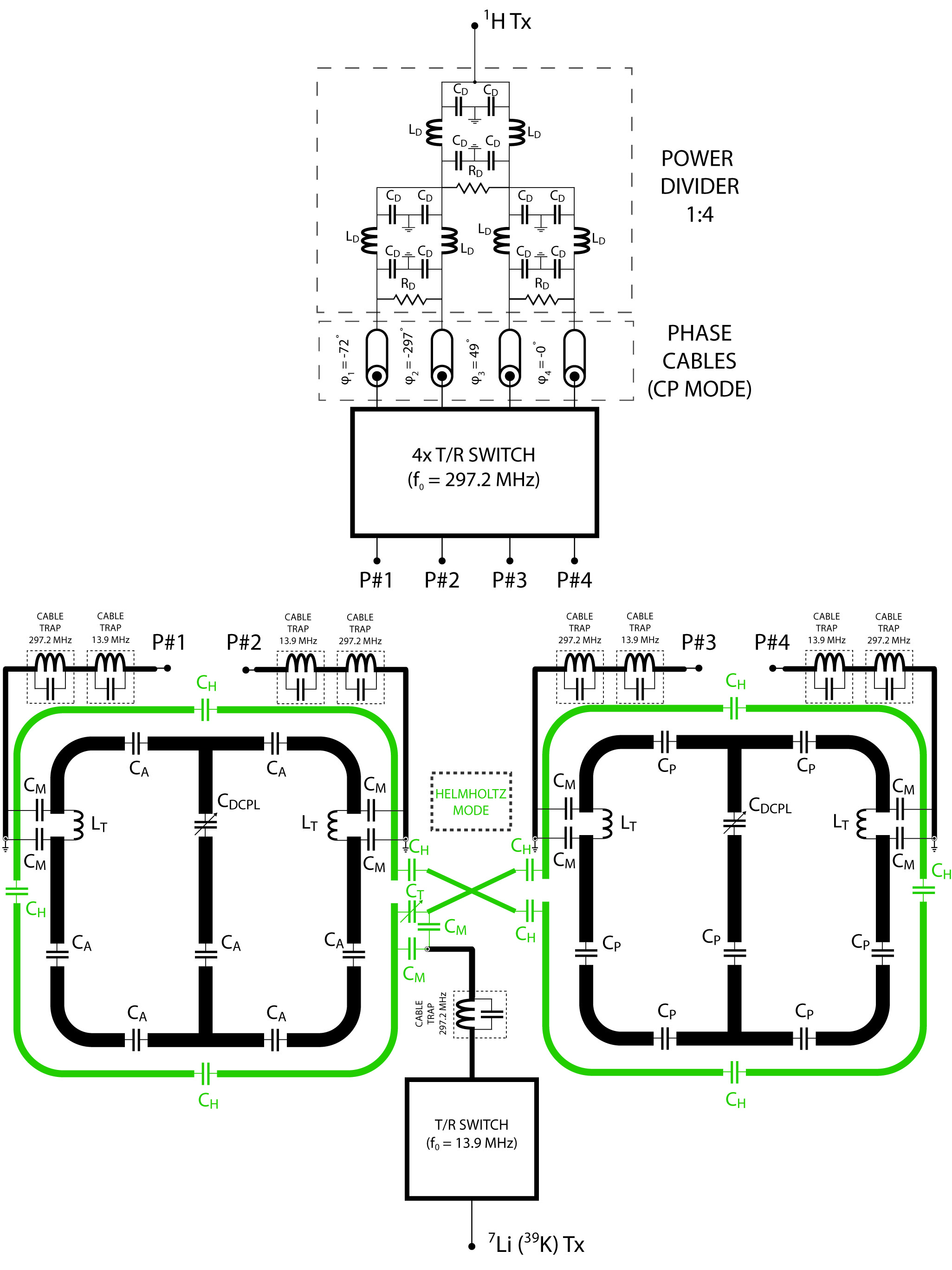
The proposed RF coil comprises two separate coils which operate at two resonant frequencies: f39K = 13.9 MHz and f1H = 297.2 MHz. It consists of two sections: anterior (curved to conform to an average human torso) and posterior (planar to fit into the patient table) as demonstrated in Figure 1. Each section is composed of one large loop element (270x280mm2) which was tuned to f39K, and - concentrically - two smaller loop elements (220x200mm2) which were tuned to f1H (Figure 2). The 39K loop elements were connected to support the Helmholtz mode. The 1H loop elements were driven in a circularly polarized (CP) mode. Electromagnetic field (EMF) and specific absorption rate (SAR) simulations were performed using CST Studio Suite 2015 (CST AG, Darmstadt, Germany) involving the human voxel models Duke and Ella. Phantom and human imaging studies were conducted on a 7.0 T whole-body MR system (Magnetom, Siemens Healthineers, Erlangen, Germany). 39K MRI was performed using previously demonstrated approach4,5.Results
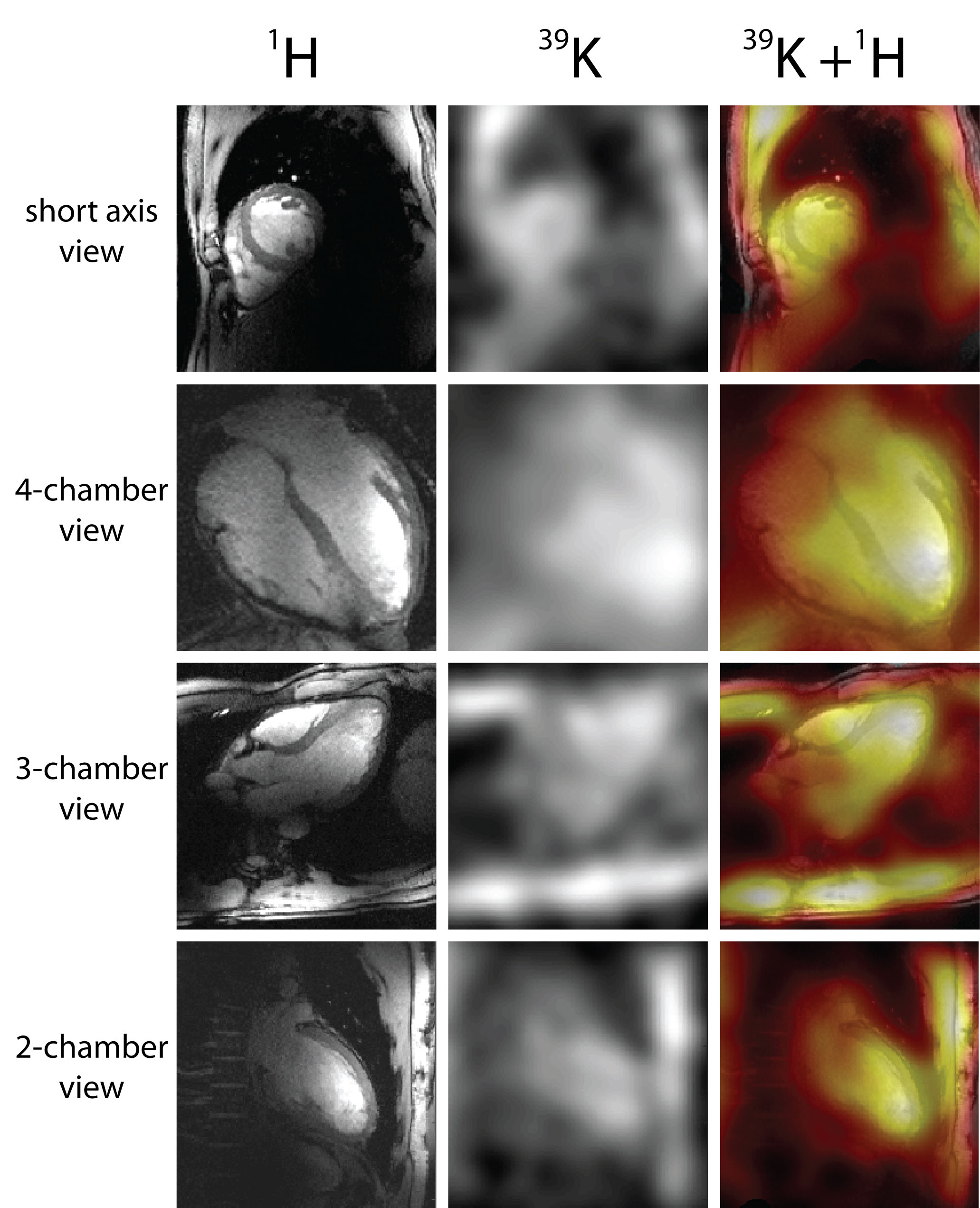
The coupling between the 39K coil and the four-channel 1H array was investigated. It was found to be negligible at 13.9 MHz (all below -33.4 dB). Coupling at 297.2 MHz is in the acceptable range between -21.8 dB and – 33.0 dB. The QUL/QL ratio for the 39K Helmholtz coil was QUL/QL = 5.2 (QUL = 141, QL = 27), To achieve the CP mode within the cardiac ROI in the human voxel model Duke the phases were set to: channel 1: -72°, channel 2: -297°, channel 3: -49°, channel 4: 0°. The same phase setting was applied for the human voxel Ella (Figure 3). Local SAR values averaged over 10 g (SAR10g) were derived from the EMF simulations for both frequencies using the human voxel models Duke and Ella for an input power of 1 W (Figure 4). Local SAR maxima were 0.21 W/kg for Duke and 0.22 W/kg for Ella for f39K and 0.46 W/kg for Duke and 0.46 W/kg for Ella for f1H. The in vivo feasibility study provided 39K images along with 1H images of the human heart which are highlighted in Figure 5. For 39K MRI of the heart, a nominal isotropic spatial resolution of (14.5x14.5x14.5) mm3 was achieved within 30 minutes scan time.Discussion and Conclusion
In this work, we demonstrated for the first time the feasibility of in vivo 39K MRI of human heart at 7.0 T. The pioneering work by Parrish et al.6 suggested that 39K MRI of the heart with isotropic spatial resolution of 13 mm3 and scan time of 30 minutes should be “clinically useful”. Their simulations revealed that the sensitivity obtained at 1.5 T was not sufficient to achieve this goal. The simulations requested an 8-fold increase in SNR which we approached with our experiments at 7.0 T. Potassium ions are crucial for life and their significance in the heart physiology and metabolism is beyond the function they play in the activity of Na+/K+-ATPase. The link between molecular mechanisms involving K+ channels expressed in human heart and higher risk of arrhythmias remains poorly understood7. The pathologic alterations related to K+ concentration reveal that these observations might have clinical importance in a number of contexts. The broad roles of this element in processes related to cardiac physiology suggest a range of questions for cardiovascular investigations.Acknowledgements
This work was funded by the Helmholtz Alliance iMED, the Helmholtz Initiative on Personalized MedicineReferences
1. Doyle DA, et al. Science. 1998;280:69-77.
2. Skou JC. Biochim Biophys Acta. 1957;23:394-401.
3. Rooney WD, Springer CS, Jr. NMR Biomed. 1991;4:209-226.
4. Nagel AM, et al. Magn Reson Med. 2009;62:1565-1573.
5. Umathum R, et al. Radiology. 2013;269:569-576.
6. Parrish TB, et al. Magn Reson Med. 1997;38:653-661.
7. Giudicessi JR, Ackerman MJ. Nat Rev Cardiol. 2012;9:319-332.
Figures