2030
Novel phase-sensitive late gadolinium enhancement MRI for assessment of myocardial infarction in large animals1Radiology, University of Washington, Seattle, WA, United States, 2Pathology, University of Washington, Seattle, WA, United States, 3Institute for Stem Cells and Regenerative Medicine, University of Washington, Seattle, WA, United States, 4Comparative Medicine, University of Washiington, Seattle, WA, United States
Synopsis
Novel phase sensitive reconstruction method of the gadolinium delayed hyperenhancement, 3D-TRIPS, was used for the first time in the infarct visualization and quantitation in the hearts of non-human primates and Yucatan mini-pigs. Advantages of using 3D-TRIPS reconstruction vs. standard 2D-PSIR method for MR imaging of infarct include scan time shortening due to elimination the background phase-reference scan and improvement of the contrast ratio between normal and infarcted myocardium as well as between scar and blood. The novel cardiac MRI 3D-TRIPS method provides robust infarct size measurements while preserving scan time.
Introduction
T1-weighted inversion-recovery (IR) magnetic resonance imaging (MRI) acquisitions are commonly used in detection of myocardial scar after contrast agent administration. Phase-sensitive detection has been used to improve infarct to myocardium contrast by preserving the sign of the desired magnetization during IR and reducing the dependence on prescribing at exact inversion time (TI). Phase-sensitive inversion recovery (PSIR) is the standard MRI method for scar imaging 1. However, phase-sensitive reconstruction requires acquisition of an additional phase-reference image which doubles the total scan time, thereby limiting its applications. A new method for imaging of myocardial scar has been recently developed 2. The 3D True Polarity Recovery with Independent Phase Estimation Using Multi-layer Stacks Based Region-Growing (3D-TRIPS) technique allows shortening imaging time by direct reconstruction of phase sensitive images without the need for a separate reference scan. Equivalent image quality for 3D-TRIPS and PSIR acquisitions has been demonstrated on patients with nonischemic cardiomyopathy 2. Shortening of scan time is important especially in complicated cases where prolonged anesthesia cannot be tolerated.
Aim of this study was to evaluate 3D-TRIPS method in imaging of myocardial infarction in the large laboratory animals typically used for pre-clinical evaluations of therapeutic interventions.
Methods
Myocardial infarction was induced in 10 Macaca nemestrina and in 15 Yucatan mini-pigs by mid-LAD balloon inflation (3 hours in monkeys; 90 minutes in pigs) followed by reperfusion. Infarct size was evaluated noninvasively with MRI in monkeys using 3T Achieva Philips scanner (Best, Netherlands) and Flex-M/Flex-L coils; pig hearts were imaged using 3T Ingenia Philips scanner with dStream torso coil.
Inversion-recovery (IR) images were acquired ~5 minutes after intravenous injection of 0.2 mmol/kg Gd-DTPA ProHance (Bracco Diagnostics Inc., Princeton, NJ). IR sequence parameters for monkey’s scan included: TI ~ 180 ms; FOV 150x150x48 mm3, slice thickness 4 mm no gap, TR/TE 7.0 /3.5 ms, FA 25°, 3 signal averages, acquired voxel size 1.2/1.2/4 mm reconstructed to 0.63/0.63/2 mm3. Scan time 5.38 min.
PSIR images were acquired ~10 minutes after contrast agent administration with following parameters: TI 280-350 ms; FOV 150x150 mm2, 12 slices, slice thickness 4 mm, TR/TE 7.0/3.5 ms, FA 25°, PSIR flip angle 5°, 3 signal averages, scan time ~8.36 min. Imaging parameters for pig’s cardiac scan were kept the same except increased FOV to 250x250 mm and increased slice thickness to 6 mm. Prospective ECG triggering was used without breath hold.
PSIR images were reconstructed with the Philips IntelliSpace Portal software. 3D-TRIPS reconstruction was conducted from the IR images with the custom-written software in Matlab 2. Infarct size was measured as percentage of the enhanced area to total left ventricle (LV) area with the threshold set as full-width-half-max. Signal intensities of the infarcted and non-infarcted areas of LV as well as inside of LV chamber (blood) were measured. Signal intensity ratios (SIR) were calculated between infarcted and non-infarcted area as well as between infarct and blood. Signal-to-noise (SNR) and contrast-to-noise (CNR) were measured in all images.
Results
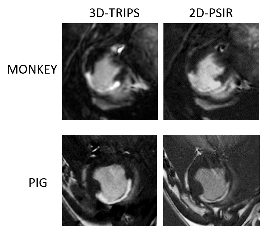
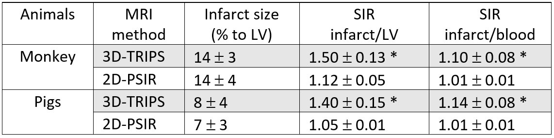
Our results have shown advantages of using 3D-TRIPS reconstruction vs. standard 2D-PSIR method for MR imaging of infarct. First, the acquisition time without acquiring phase reference image was reduced by 1.5-fold. Second, the SIR between infarcted and non-infarcted LV wall was significantly higher in the 3D-TRIPS images, 1.50±0.13, compared to 1.12±0.05 in PSIR on monkey’s heart (p<0.001, means ± St. Deviation) and 1.40 ± 0.15 in 3D-TRIPS images and 1.05 ± 0.01 in PSIR pig’s heart (p<0.001). PSIR images often suffer from the poor contrast between infarcted tissue and blood that complicates the scar delineation. The third advantage of 3D-TRIPS was higher SIR between scar and blood, 1.10±0.08 in 3D-TRIPS and 1.01±0.01 in PSIR on monkey’s heart (p<0.001); 1.14 ± 0.08 in 3D-TRIPS and 1.01 ± 0.01 in PSIR on pig’s heart (p<0.001), which enabled better visualization of endocardium and differentiation from blood (Figure 1). Infarct size measurements between 3D-TRIPS and PSIR were similar (Table 1). All images were characterized with high SNR and CNR.Discussion and conclusion
Scan time shortening might be critical for patients and animals with severe heart damage. Recently developed 3D-TRIPS reconstruction method shortens scan time by 1.5-fold due to elimination the background phase-reference scan. PSIR images often suffer from the poor contrast between infarcted tissue and blood that complicates the scar delineation. The 3D-TRIPS method improves the contrast (ratio) between normal and infarcted myocardium (scar) as well as between scar and blood. Thus, novel 3D-TRIPS method provides robust infarct size measurements while preserving scan time.Acknowledgements
No acknowledgement found.References
1. Kellman P, Arai AE, McVeigh ER, Aletras AH. Phasesensitive inversion recovery for detecting myocardial infarction using gadolinium-delayed hyperenhancement. Magn Reson Med. 2002;47(2):372–383.
2. Liu H, Wilson GJ, Balu N, et al. 3D true‑phase polarity recovery with independent phase estimation using three‑tier stacks based region growing (3D‑TRIPS). Magn Res Mater Phy. 2018;31(1):87-99.
Figures