2008
Genotypic Groups as Risk Factor for Cardiac MR Abnormalities and Complications in Thalassemia Major1Fondazione G. Monasterio CNR-Regione Toscana, Pisa, Italy, 2Fondazione di Ricerca e Cura "Giovanni Paolo II", Campobasso, Italy, 3Ospedale "Sandro Pertini", Roma, Italy, 4"ARNAS" Civico, Di Cristina Benfratelli, Palermo, Italy, 5Ospedale “SS. Annunziata” ASL Taranto, Taranto, Italy, 6Azienda Ospedaliera "Garibaldi", Presidio Ospedaliero Nesima, Catania, Italy, 7Policlinico di Modena, Modena, Italy, 8P.O. Pediatrico Microcitemico “A.CAO”, Cagliari, Italy, 9Ospedale "V. Emanuele III", Gela (CL), Italy
Synopsis
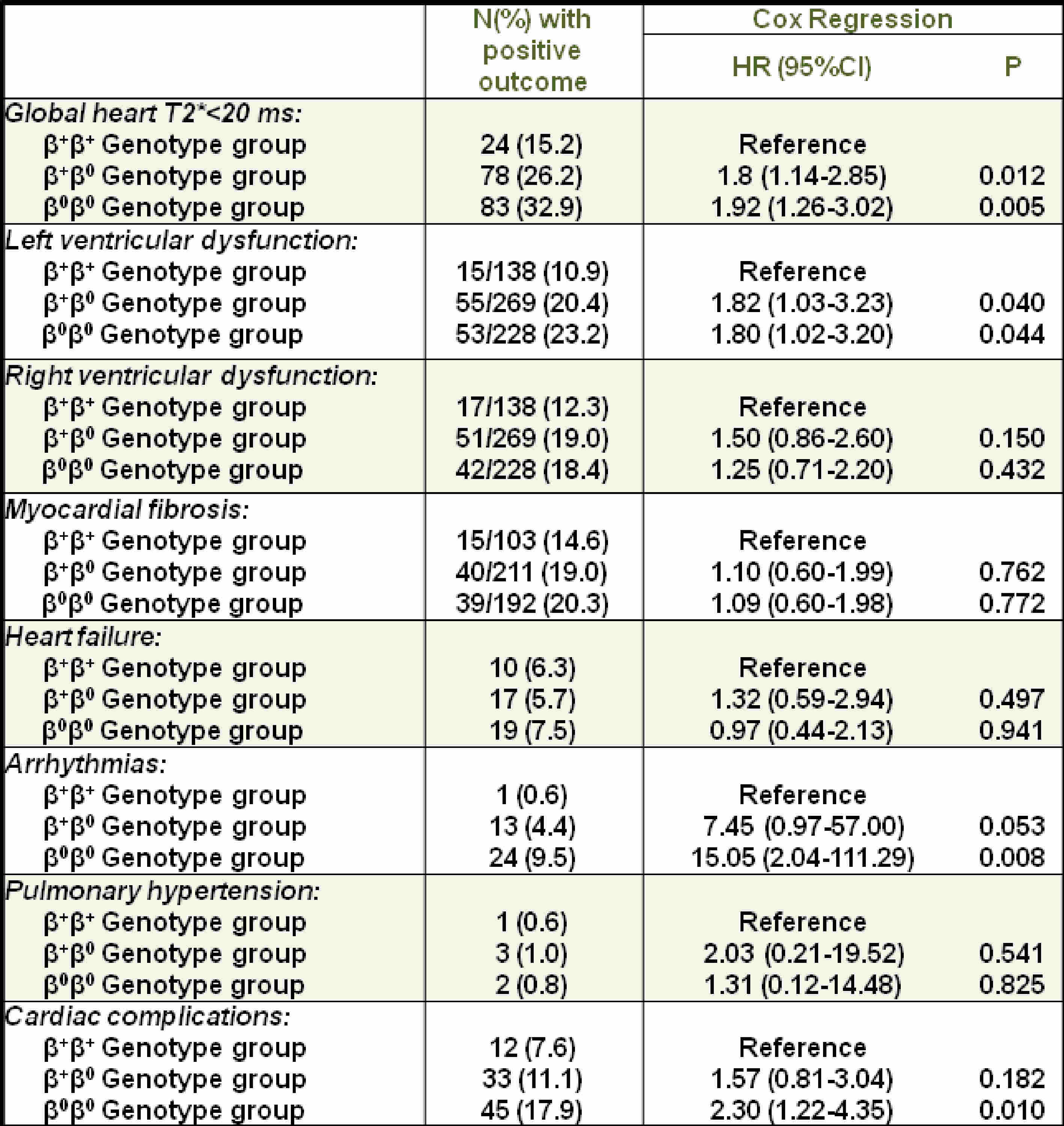
On the basis of the type of gene mutation, three groups of patients with thalassemia major (TM) were identified: homozygotes β+, compound heterozygotes β+/β° and homozygotes β°. Compared to the milder genotype group homozygotes β+, the other two groups showed a significantly higher risk of myocardial iron overload (MIO) and left ventricular dysfunction. Moreover, homozygotes β° showed a significantly higher risk of CC than homozygotes β+ patients. These data support the knowledge of the different genotypic groups in the clinical management of β-TM patients.
Introduction
Beta thalassemia major (β-TM) displays a great deal of genotypic heterogeneity, not fully investigated in terms of cause-effect1.
This prospective and multicentre study aimed to detect if different genotypic groups could predict the development of cardiovascular magnetic resonance (CMR) abnormalities and cardiac complications (CC).
Methods
We considered 708 β-TM patients (373 females, 30.05±9.47 years), consecutively enrolled in Myocardial Iron Overload in Thalassemia (MIOT) network2. Data were collected from birth to the first CMR imaging scan.
Myocardial iron overload was assessed by the multislice multiecho T2* technique3. Biventricular function parameters were quantified by cine images4. Late gadolinium enhancement (LGE) images were acquired to detect myocardial fibrosis5.
Results
On the basis of the type of gene mutation, three groups of patients were identified: homozygotes β+ (N=158), compound heterozygotes β+ / β° (N=298) and homozygotes β° (N=252).
Table 1 shows the effect of genotype group on the development of different cardiac outcomes. Compared to the milder genotype group homozygotes β+, the other two groups showed a significantly higher risk of myocardial iron overload (MIO) and left ventricular dysfunction.
We recorded 90 (13.0 %) cardiac events: 46 heart failures (HF), 38 arrhythmias (33 supraventricular, 3 ventricular and 2 hypoinetic) and 6 pulmonary hypertensions (PH). No prospective association was detected between genotype group and HF and PH. The homozygous β° group showed a significantly higher risk of arrhythmias than the homozygous β+ group and at the limit of significance than the compound heterozygotes. Globally, homozygotes β° showed a significantly higher risk of CC than homozygotes β+.
Conclusions
Different genotypic groups predict the development of MIO, left ventricular dysfunction, arrhythmias and CC in β-TM patients. These data support the knowledge of the different genotypic groups in the clinical management of β-TM patients.Acknowledgements
No acknowledgement found.References
1. Thein SL. The molecular basis of beta-thalassemia. Cold Spring Harb Perspect Med 2013;3(5):a011700.
2. Meloni A, Ramazzotti A, Positano V, et al. Evaluation of a web-based network for reproducible T2* MRI assessment of iron overload in thalassemia. Int J Med Inform 2009;78(8):503-512.
3. Meloni A, Positano V, Keilberg P, et al. Are the Preferential Patterns of Myocardial Iron Overload Preserved at the Cmr Follow-Up? Journal of Cardiovascular Magnetic Resonance 2012;14(Suppl 1):190.
4. Aquaro GD, Camastra G, Monti L, et al. Reference values of cardiac volumes, dimensions, and new functional parameters by MR: A multicenter, multivendor study. J Magn Reson Imaging 2016;45(4):1055-1067.
5. Pepe A, Positano V, Capra M, et al. Myocardial scarring by delayed enhancement cardiovascular magnetic resonance in thalassaemia major. Heart 2009;95:1688-1693.
Figures