1993
Vortex Formation Time in Chinese Children: a CMR Study1Radiology, Shanghai Children's Medical Center, Shanghai, China, 2Shanghai Children's Medical Center, Shanghai, China, 3MR Research GE Healthcare, Shanghai, China, 4Radiology, University of Wisconsin-Madison, Madison, WI, United States
Synopsis
Vortex formation time (VFT) is an index of
left ventricular (LV) systolic and diastolic performance, with normal values,
based on echocardiography, from 3.3 to 5.5 ms in adults [1]. With the
increasing intensity of vortex, the vortex ring is pinched off; this instant is
defined as the vortex ring formation time. With echocardiography VFT is
measured from trans-mitral inflow velocities. Although cardiovascular magnetic
resonance (CMR) is the gold standard for assessing LV systolic function, its use
in evaluating LV diastolic function is more limited [2]. Through-plane motion of
the mitral valve results in underestimations of the peak
mitral inflow velocities with standard 2D flow CMR acquisitions. 4D flow
CMR analysis improves the accuracy of peak mitral inflow velocities because of
its ability to track mitral valve plane motion [3]. However, vortex ring
quantification with 4D flow CMR has not been widely used [4]. The VFT has not
been evaluated in children and could be useful tools for assessment of
diastolic function using CMR.
Purpose
The aim of this study was to quantify VFT and establish normal pediatric VFT values using 4D flow CMR in children ages 6-18 years.Methods
Subjects: 4D flow CMR data from 46 healthy Chinese children (6-18 years, M:31; Female:15) were retrospectively identified from studies performed between June 2017 - August 2018. All volunteers had normal blood pressure and were free of cardiovascular disease. This case-control study was approved by our institutional review board and research images was performed after signed informed consent.
CMR imaging: CMR was conducted on a 3.0 Tesla scanner (Discovery MR 750, GE, USA) using the 8-channel phased-array cardiac coil. Whole heart 4D flow CMR was performed (spatial resolution=1.2-1.8×.1.2-1.8×1.2-1.8 mm3, FOV=340-420 mm2, slab thickness=60-85 mm, temporal resolution=34.4 ms, TE=2.1 ms, TR=4.3 ms, flip angle=8-12°, bandwidth=62.5 Hz/pixel, velocity sensitivity=160cm/s) with a coronal or sagittal slab acquisition covering the entire heart. The scan time was 7-11 min. No respiratory gating and contrast agent were used in the study. Three-chamber and short-axis cine balanced steady state free precession images were also acquired using standard clinical protocols.
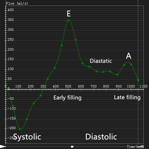
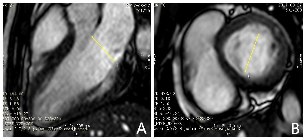
CMR analysis: CMR analysis was performed with commercially available software (Cvi42, Circle Cardiovascular Imaging, Canada). The mass, EDV, ESV, SV and EF of the LV were determined from short-axis cine images. Papillary muscles and trabeculations were included with the LV blood pool. The trans-mitral valve velocities and time to peak of E-wave and A-wave were determined from the 4D flow CMR data (Figure 1). The vortex formation time(VFT) was calculated from the following equation: VFT=4/π⋅α3⋅EF⋅EWV/SV=4/π⋅EWV/D3 where α was defined as:α=EDV(1⁄3)/D, with the diameter of the mitral valve(D) calculated as the mean of maximum distance between the leaflet tips during the rapid filling phase, perpendicular to the flow direction, in the three-chamber view, and the distance between the commissures on a short-axis slice through the mitral valve (Figure 2). The E-wave volume (EWV) was defined as the difference in volumes of the LV at the end of the E-wave and ESV [5].
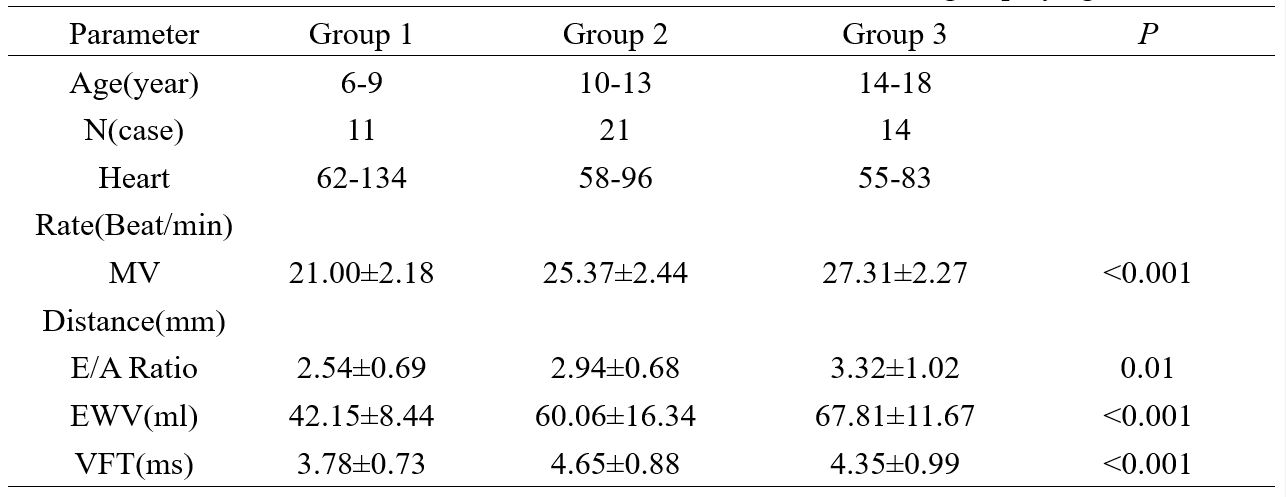
Statistical analysis: Data were categorized into three age groups (Group 1: 6-9 Years; Group 2: 10-13 Years; Group 3: 14-18 Years). Comparisons among these groups were made with one-way ANOVA analysis for normally distributed data and Kruskal-Wallis test for non-normally distributed continuous variable. Statistical significance was indicated by p<0.05.
Results
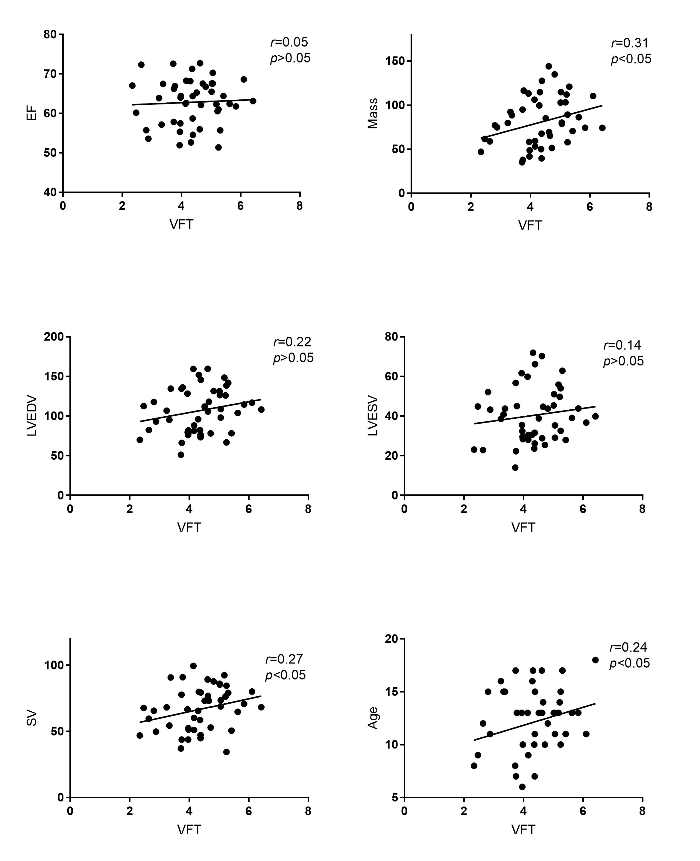
The VFT was significantly correlated with age, SV and cardiac mass (Figure 3). E/A, EWV and mitral valve diameters increased with age. The normal range of VFT is from 2.34 to 6.42 in Chinese children (Table 1). VFT in group 1 was significantly lower than VFT in groups 2 and 3 (p<0.05). The differences in VFT in groups 2 and 3 were not significant (p>0.05).
Conclusions
VFT was calculated in 46 healthy Chinese children. Normative values were determined from children ages 6-18 years. VFT could provide additional information on diastolic function in patients with congenital heart disease using 4D flow CMR.Acknowledgements
No acknowledgement found.References
- Pasipoularides A, Vlachos P P, Little W C. Vortex formation time is not an index of ventricular function [J]. Journal of Cardiovascular Translational Research, 2015, 8(1):54-58.
- Panesar D K, Burch M. Assessment of Diastolic Function in Congenital Heart Disease:[J]. Frontiers in Cardiovascular Medicine, 2017, 4(15):5.
- Kamphuis VP, Roest AAW, Ajmone Marsan N, et al. Automated Cardiac Valve Tracking for Flow Quantification with Four-dimensional Flow MRI[J]. Radiology. 2018 Oct 30:180807.
- Gharib M, Rambod E, Kheradvar A, et al. Optimal vortex formation as an index of cardiac health[J]. Proceedings of the National Academy of Sciences of the United States of America, 2006, 103(16):6305-6308.
- Töger J, Kanski M, Carlsson M, et al.
Vortex Ring Formation in the Left Ventricle of the Heart: Analysis by 4D Flow
MRI and Lagrangian Coherent Structures[J]. Annals of Biomedical Engineering,
2012, 40(12):2652-2662.
Figures