1991
Non-invasive estimations of turbulence driven relative pressure drops – applying the concept of virtual fields on 4D flow MRI1Department of Biomedical Engineering and Health Systems, KTH Royal Institute of Technology, Stockholm, Sweden, 2Clinical Sciences, Karolinska Institutet, Stockholm, Sweden, 3Department of Mechanical and Biomedical Engineering, Kangwon National University, Chuncheon, Korea, Republic of, 4Center for Medical Image Science and Visualization (CMIV), Linköping University, Linköping, Sweden, 5Division of Imaging Sciences and Biomedical Engineering, King's College London, London, United Kingdom, 6Departments Biomedical Engineering and Cardiac Surgery, University of Michigan, Ann Arbor, MI, United States
Synopsis
4D flow MRI with six-directional flow encoding has enabled the assessment of turbulent flows, including mapping of incoherent flow variance. Using such, non-invasive estimation of turbulence-driven pressure drops can be computed. Here, we present an extension of the virtual-Work-Energy-Relative-Pressure method8 for the assessment of turbulence-driven pressure drops. Using the concept of virtual fields, the method accurately assesses pressure drops over a range of stenotic valve phantoms, being validated against catheter-based measurements. With virtual probing enabling the assessment of pressure drops through complex, narrow vasculatures, the incorporation of turbulence enhances the utility of the method, enabling for refined clinical hemodynamic analysis.
Introduction
Changes in regional blood pressure have shown to correlate to a range of cardiovascular pathologies1-2. Specifically, many cases of clinical interest, such as aortic stenosis, have been shown to involve pressure changes originating from turbulent, incoherent flow3. With the development of 4D flow MRI with six-directional flow encoding (ICOSA6), a direct quantification of turbulence production is enabled4. Methods have been proposed to derive turbulence-driven irreversible pressure drops from 4D flow data3-6. The development of these techniques is however still in an exploratory phase where complex anatomies and clinical noise levels might impede method accuracy. Recently, we have shown that using the concept of work-energy7 and virtual fields8, pressure drops through narrow and bifurcating vasculatures can be assessed with high accuracy even under clinical noise and sampling conditions8. To broaden the applicability, we here present an extension to this method, incorporating a turbulence-derived energy component into the virtual work-energy relative pressure estimation.Methods
The proposed method originates from a work-energy formulation (vWERP) assessing pressure drops from a flow field $$$v$$$7 using an additional virtual field $$$w$$$ to probe any vascular segment within the imaged domain8. By doing so, we have shown that the influence of measurement inaccuracies, low flow magnitudes and flow bifurcations can be minimized8.
To extend the method to handle turbulent flow, flow inconsistencies are introduced in the original formulation6, where such is given by the variance in the flow assessed by ICOSA6 4D flow MRI. With such, and following our previously proposed derivation of virtual energy7, the pressure drop $$$\Delta p$$$ over any given vascular segment can be evaluated by:
$$\Delta p=-\frac{1}{Q_w} (\frac{\partial}{\partial t}K_w+A_w+V_w+T_w)$$
with $$$K_w$$$, $$$A_w$$$ and $$$V_w$$$ being the kinetic, advective, and viscous virtual energy components when using $$$w$$$ as the weighting velocity field, and $$$Q_w$$$ being the flow of $$$w$$$ over the domain inlet (for details see6,7). Additionally, $$$T_w$$$ is the turbulent energy component of $$$v$$$ with $$$w$$$ as the weighting velocity field, with the term originating from the assessed variance of $$$v$$$ by:
$$T_w=-\rho \int_{\Omega} Cov\left[v,v\right] \cdot \triangledown w \cdot dx$$
with $$$\rho$$$ being the fluid density, $$$\Omega$$$ the domain over which the flow is being assessed, and $$$Cov\left[v,v\right]$$$ the covariance matrix of the observed flow.
To evaluate method performance, an in-vitro setup from5 was used. ICOSA6 4D flow MRI (for a full methodolgical description, see5) was acquired for 7 different shaped stenotic valve phantoms at 3 different flow speeds. Irreversible pressure drops were assessed over all valves and compared against invasive catheter measurements. Estimates were also compared against alternative measures for turbulence-driven pressure drop assessment, evaluated in previous work5.
Results
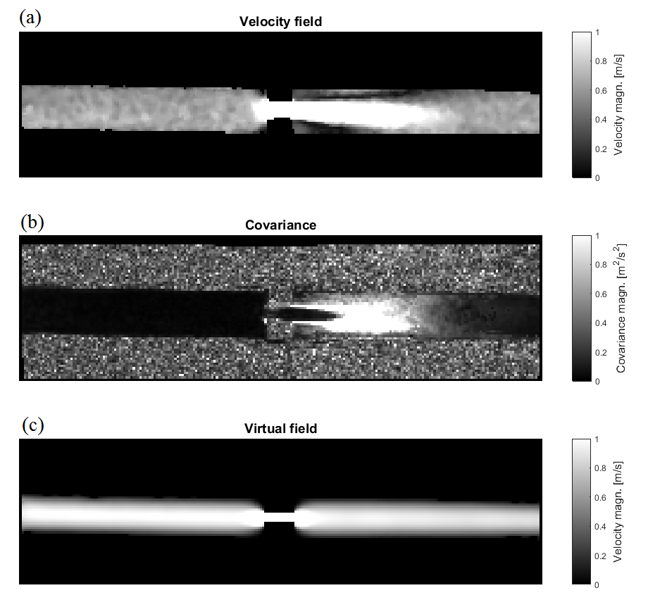
Figure 1 shows the imaged velocity field, estimated flow variance, and virtual field for one of the imaged phantom valves. With the virtual field solved as a Stokes flow boundary value problem, the field is primarily governed by the geometry of the phantom, with maximum velocities in and around the stenosis.
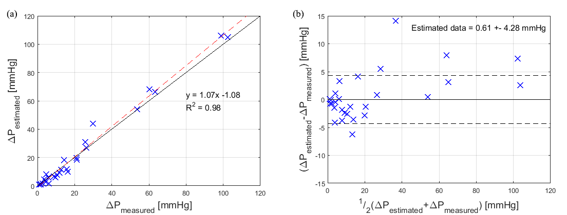
Figure 2 shows summarized results. As seen, all pressure drops were assessed at high accuracy, with only a slight overestimation of 0.6 mmHg (Figure 2b), with the overestimation seemingly increasing with increasing pressure drop magnitude (Figure 2a). The linear regression coefficient of 0.98 (Figure 2a) indicates a well-tuned assessment method, with estimated values coinciding well with the invasively measured pressure drops.
The proposed method using virtual fields also compared well when evaluated against other methods tested on the same phantom dataset (see5). In particular, the method seems to render results with lower variance, indicating increased precision with maintained accuracy compared to previously published methods.
Discussion
The correspondence between invasive catheter measurements and non-invasive 4D flow MRI-based estimates demonstrates a good accuracy of the vWERP method. In particular, vWERP showed to be able to obtain accurate pressure drop estimates over a range of different turbulence levels and valve configurations. With the original vWERP formulation shown to enable dynamic probing of pressure drops through complex, narrow, bifurcating cardiovascular structures8, this turbulent energy extension would widen the applicability of the method and would potentially enable a complete assessment of relative pressure through any imaged cardiovascular structure. Detailed evaluations of the method’s spatiotemporal behavior and in-vivo performance in the setting of turbulent flow would further clarify the range and applicability of the method.Conclusion
We show that turbulence-driven hemodynamic pressure drops can be successfully assessed from 4D flow MRI using the concept of virtual fields after extending the vWERP method by introducing a turbulent energy term. In particular, by extending a method already shown viable in complex, bifurcating vascular structures, the clinical applicability of this method is even further enlarged.Acknowledgements
We would like to acknowledge funding from Engineering and Physical Sciences Research Council (EP/N011554/1). This work was also supported by the Wellcome EPSRC Centre for Medical Engineering at King’s College London (WT 203148/Z/16/Z).References
1. Baumgartner H, Hung J, Bermejo J, Chambers JB, Evangelista A, et al. Echocardiographic assessment of valve stenosis: EAE/ASE recommendations for clinical practice. J. Am. Soc. Echocardiogr. 2008:101-102
2. Vahanian A, Baumgartner H, Bax J, Butchart E, Dion R, et al. Guidelines on the management of valvular heart disease: the task force on the management of valvular heart disease of the european society of cardiology. Eur. Heart J. 2007:28(2):230-68
3. Dyverfeldt P, Hope MD, Tseng EE, Saloner D, Magnetic Resonance measurement of turbulent kinetic energy for the estimation of irreversible pressure loss in aortic stenosis. JACC: Cardiovasc Im. 2013:6(1):64-71
4. Ha H, Lantz J, Ziegler M, et al. Estimating the irreversible pressure drop across a stenosis by quantifying turbulence production using 4D flow MRI. Sci Rep. 2017:7:46618
5. Ha H, Escobar Kvitting J-P, Dyverfeldt , Ebbers T. Validation of pressure drop assessment using 4D flow MRI-based turbulence production in various shapes of aortic stenosis. Magn Res Med. 2018:1-14
6. Gülan U, Binter C, Kozerke S, Holzner M. Shear-scaling-based approach for irreversible enery loss estimation in stenotic aortic flow – an in vitro study. J Biomech. 2017:56:89-96
7. Donati F, Figueroa CA, Smith NP, Lamata P, Nordsletten DA. Non-invasive pressure difference estimation from PC-MRI using the work-energy equation. Med. Image Anal. 2015:26(1):159-72
8. Marlevi D, Ruijsink B, Balmus M, Dillon-Murphy D, Fovargue D et al. Non-invasive pressure estimations by virtual fields – cardiovascular pressure drops from 4D flow MRI. ISMRM 2018, Paris, France.
Figures