1953
Dual-Venc 4D Spiral Imaging of Aortic Flows1Electrical and Computer Engineering, University of Louisville, Louisville, KY, United States, 2Robley Rex VA Medical Center, Louisville, KY, United States, 3Philips Health Care, Gainesville, FL, United States, 4Division of Cardiovascular Medicine, University of Louisville, Louisville, KY, United States
Synopsis
Dual-Venc is a technique for MR flow imaging which uses two Vencs to acquire a cardiac cycle, improving diastolic data. Dual Venc 4D flow with spiral readouts was used to image the outflow tract and through the aortic valve in both phantom and patients with severe aortic stenosis. In-vitro model of the aortic arch included a calcific polymeric valve which behaved physiologically. The results of in-vitro and in-vivo scans show that 4D Spiral Dual-Venc Flow is comparable in results to 4D Cartesian Flow in systole, while improving diastolic data, and reducing scan time by 30% to 50%.
Introduction
Calcification of the aortic valve is the most common cause of a chronic and progressive valvular disease called aortic stenosis (AS) which if left untreated has a 50% 2-year survival rate [1]. While Doppler US is the initial diagnostic tool in evaluating patients with AS, it is limited by availability of adequate acoustic windows and the technical expertise of the sonographer. MRI is a true 3D modality which may provide additional advantages over ultrasound in the detection and evolution of AS in certain patient populations.Methods
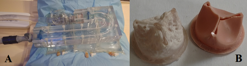
Scans were performed on a Philips Achieva 1.5 T with a 16 channel SENSE XL Torso coil. The proposed dual Venc sequence was compared with a single Venc sequence with Cartesian read out. The end systolic time point was used as the switch time for transitioning from a high Venc acquisition to a low Venc. The aortic arch phantom including the valve [3] used in the in-vitro studies can be seen in Figure 1.
The phantom which was machined from clear acrylic was connected to a programmable physiologic pump on the inlet, and a fluid reservoir on the outlet via plastic tubing. The circulating fluid was comprised of 60% distilled water and 40% glycerol, which results in viscosity = 0.0043 Pascal*s, and density = 1060 kg/m3. The dual Venc sequence with systolic/diastolic Vencs350/150 cm/s were applied in conjunction with Cartesian and Spiral readout trajectories. The field of view was 100mm x 100mm x 48mm, with a voxel size of 1.5mm x 1.5mm x 3 mm, 16 slices with coverage from proximal to the valve to 27mm distal to the valve, and 28 phases. Other scan parameters were as follows: TR=14 ms, TE=4.2 ms (Cartesian), and TE=1.75ms (Spiral). Number of spiral interleaves = 32, length of readout = 4ms, and TFE factor=1 for both acquisition types [2]. For the in-vivo acquisitions, FOV= 200 mm x 200 mm x 50 mm resulted in a resolution of 2.5 x 2.5 mm in –plane and a slice thickness of 5 mm. This was acquired over 16 heart phases, with a flip angle of 8 degrees. The Venc used was defaulted to 400 cm/s for systole, and 100 cm/s for diastole. Any changes to the Venc were determined from a 2D PC-MRI through-plane acquisition performed immediately before the 4D Flow acquisitions. The rest of the scan parameters were similar to the in-vitro acquisition. Data were obtained in 6 healthy volunteers, and 7 patients with severe AS.
Results
The flow waveforms were calculated for all acquisition types and plotted in a scatter plot to directly compare net flows from different acquisitions against each other. From the scatter plot the Pearson correlation coefficient (p<0.01) was determined [4].
The flow waveform results can be seen in Figure 2a, Figure 3a, and Figure 4a. Figure 2a shows flow at slice 10 distal to the polymeric valve for the 0%, calcific and 50% calcific valve. Figure 3a is the flow waveform for a healthy volunteer, and Figure 4a is for the AS patient. The flow waveform results (Spiral Dual Venc and Cartesian single Venc) are in agreement. The scatter plot comparison of the net flows can be seen in Figure 2b, Figure 3b, and Figure 4b. Figure 2b is the phantom scatter plot, with the 0% valve having a Pearson correlation of 0.983, while the 50% coefficient of 0.990. The healthy volunteer, Figure 3b, correlation coefficient is 0.989, while the patient, Figure 4b, has a correlation coefficient of 0.977. There is some expectation that the correlation coefficients would decrease as the flow got more complex, however the exception to this is the 0% phantom results. Figure 2c/d, Figure 3c/d, and Figure 4c/d show the through-plane velocity distribution for the same slice position that was used to compute the flow waveform at peak systolic time. Velocities appear similar, with the Cartesian acquisition having larger peak velocities.
Conclusion
This study shows that 4D Dual Venc Spiral Flow MRI results in similar flow waveforms to 4D Cartesian Flow counterpart for in-vitro and in-vivo physiologic valvular flows in patients with AS. The Spiral acquisition can do this with a roughly 30% to 50% scan time reduction [2] relative to a non-accelerated 4D Flow acquisition with Cartesian readout. Owing to the lower Venc during the diastolic phase of the acquisition, 4D Dual Venc renders diastolic velocities less noisy with improved velocity resolution [3].Acknowledgements
This research has been supported by NIH Grant R21HL132263References
[1] C. M. Otto, B. K. Lind, D. W. Kitzman, B. J. Gersh and D. S. Siscovick, "Association of aortic-valve sclerosis with cardiovascular mortality and morbidity in the elderly," New England Journal of Medicine, vol. 341, pp. 142-147, 1999.
[2] A. Nilsson, K. M. Bloch, M. Carlsson, E. Heiberg and F. Ståhlberg, "Variable velocity encoding in a three-dimensional, three-directional phase contrast sequence: evaluation in phantom and volunteers," Journal of Magnetic Resonance Imaging, vol. 36, pp. 1450-1459, 2012.
[3] M. Negahdar, M. Kadbi, M. Kendrick, M. F. Stoddard and A. A. Amini, "4D spiral imaging of flows in stenotic phantoms and subjects with aortic stenosis," Magnetic resonance in medicine, vol. 75, no. 3, pp. 1018--1029, 2016.
[4] S. Schnell, S. A. Ansari, C. Wu, J. Garcia, I. G. Murphy, O. A. Rahman, A. A. Rahsepar, M. Aristova, J. D. Collins, J. C. Carr and M. Markl, "Accelerated dual-venc 4D flow MRI for neurovascular applications," Journal of Magnetic Resonance Imaging, 2017.
[5] A. Falahatpisheh, D. Morisawa, T. T. Toosky and A. Kheradvar, "A calcified polymeric valve for valve-in-valve applications," Journal of Biomechanics Vol.50, pp. 77--82, 2017.
Figures