1948
Fat-Water Magnetic Resonance Imaging Reveals Dynamic Alterations in Brown Adipose Tissue Lipid Content During Cold Exposure1Institute of Imaging Science, Vanderbilt University Medical Center, Nashville, TN, United States, 2Radiology and Radiological Sciences, Vanderbilt University Medical Center, Nashville, TN, United States, 3Biomedical Sciences, Grand Valley State University, Allendale, MI, United States
Synopsis
Fat-water MRI (FWMRI) provides a powerful means of exploring changes in the lipid content of brown adipose tissue (BAT) in response to cold exposure. Most previous FWMRI studies of changes in BAT's fat signal fraction (FSF) have done so only before and after cold exposure, however, and studies have used different FSF thresholds for defining BAT. Here, we show that in healthy young men, cold exposure elicits varied responses in BAT FSF, with areas of higher initial lipid content undergoing a net lipid depletion and areas of lower initial lipid content undergoing a net lipid accumulation.
Introduction
Fat-water magnetic resonance imaging (FWMRI) provides a powerful means of exploring the changes in the lipid content of brown adipose tissue (BAT) in response to cold exposure. Most previous FWMRI studies of changes in the fat signal fraction (FSF) of BAT have done so only before and after cold exposure, however, and as a result the dynamics of FSF during cold exposure remain unappreciated. Also, the FWMRI studies of BAT reported in the literature have used different FSF thresholds for defining BAT. How the setting of these thresholds impacts the reporting of the FSF response to cold, and consequently our understanding of BAT’s physiology, is unclear.Purpose
To use FWMRI to assess changes in the FSF values of BAT and other organs during whole-body cooling in healthy young men and to explore how these changes may depend on the initial or thermoneutral FSF value.Methods
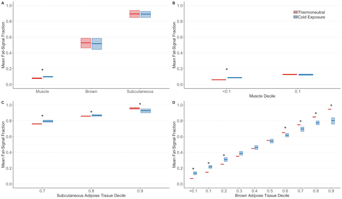
Eight, healthy men (age: 26.7 ± 3.4 years; body mass index: 23.6 ± 2.5 kg/m2) completed three visits: study familiarization, perception-based cooling (1) to identify an individualized shiver threshold (water blanket temperature: 15.6 ± 2.4 °C), and cooling during MRI. During the MRI visit, FSF maps of the supraclavicular region were acquired essentially as previously described (2). Data were acquired in a thermoneutral condition and while the subject experienced progressive cooling to 3°C above the shiver threshold temperature.
The images were registered to the initial image in the dataset using a 3D Demons algorithm. The following analyses were performed only in images that had been processed using image registration. We manually segmented the images to define regions of interest (ROI) in supra-clavicular BAT, subcutaneous adipose tissue (SAT), and lean muscle (MUS). To explore changes in FSF as a function of initial FSF content, we assigned voxels in each tissue to FSF deciles: <0.1, 0.1-0.2, … 0.9-1.0 and measured the difference in mean FSF values between thermoneutral and cold exposure.
Results
When considering all BAT voxels, mean FSF did not differ in response to cold exposure. SAT likewise underwent no net change, but there was a small increase in the observed FSF in MUS (Figure 1A). In all tissues, however, there were decile-specific changes in FSF (Figure 1, B-D). In BAT, cold exposure elicited a net increase in FSF in the lowest FSF deciles; Wilcoxon signed rank test, p<0.05) and a significant decrease in FSF in the highest FSF deciles (p<0.05) (Figure 1D). These changes occurred as linear functions of time (data not shown).Discussion
These data suggest that in healthy young men, cold exposure elicits varied responses in BAT lipid content, with areas of higher initial lipid content undergoing a net lipid depletion and areas of lower initial lipid content undergoing a net lipid accumulation.Acknowledgements
Funding: NIH R01 DK105371 & NCATS/NIH UL1 TR000445References
(1) Coolbaugh CL, et al (2018). An individualized, perception-based protocol to investigate human physiological responses to cooling. Front Physiol 9:195.
(2) Gifford A, et al. (2016). Characterizing active and inactive brown adipose tissue in adult humans using PET-CT and MR imaging. Am J Physiol Endocrinol Metab 311:E95.
Figures