1934
Regional differences of IVIM-measured perfusion fraction in vertebral bone marrow at 1.5 T.1Univ Rennes, Inserm, LTSI – UMR 1099, Rennes, France, 2CHU Rennes, Rennes, France
Synopsis
The purpose of this study was to assess regional differences of IVIM (Intra-Voxel Incoherent Motion) parameters in vertebral bone marrow. A RESOLVE Diffusion sequence with SPAIR fat suppression was acquired on healthy volunteers. The current study shows a significant difference in perfusion fraction f between the anterior and posterior regions in lumbar vertebrae L1 to L5.
Introduction
There is
in increasing interest in MRI quantitative investigations of bone marrow1. On the other hand, only a handful
of IVIM-based measurements of perfusion and diffusion have been performed to date
on vertebral bone marrow2.
In previous studies, the quantitative analysis on vertebral IVIM parameters was performed on regions of interest, whose exact location was sometimes not well specified. This approach might introduce a confounding factor in the measured IVIM parameters. Indeed, the vertebra’s posterior region is more vascularized than the anterior region; therefore, the IVIM parameters might be affected by this anatomical difference.
The purpose of this study was to determine whether the difference of vascularization between posterior and anterior region would affect the perfusion and diffusion values measured with IVIM.
Materials & Methods
MR Imaging: The lumbar spines of 6 healthy volunteers (23±4 years, 4 women and 2 men) were scanned at 1.5T (MAGNETOM Aera, Siemens Healthcare, Erlangen, Germany) using the RESOLVE diffusion-weighted sequence with TR/TE 2400/58ms, FOV 400x400mm², matrix 188x188, 10 slices of 6mm, iPAT 3, 3 segments in the readout direction, 2 averages, b-values=[0,50,100,150,400,800,1000 s/mm²] and with the SPAIR (Spectral Attenuated Inversion Recovery) fat suppression module for 4:41min of acquisition time.
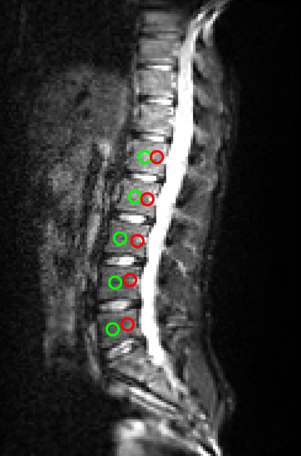
Data Analysis: Two 25-voxel-area ROIs were drawn for each lumbar vertebra (L1 to L5) on each subject with ImageJ (NIH, Bethesda, MD, http://imagej.nih.gov/ij/) [Figure1]. The mean value of the signal was measured for each b-value and fitted to the bi-exponential IVIM model. The IVIM parameters f, D and D* were determined with in-house-built scripts in Mathematica (Wolfram Research, Champaign, IL, USA).
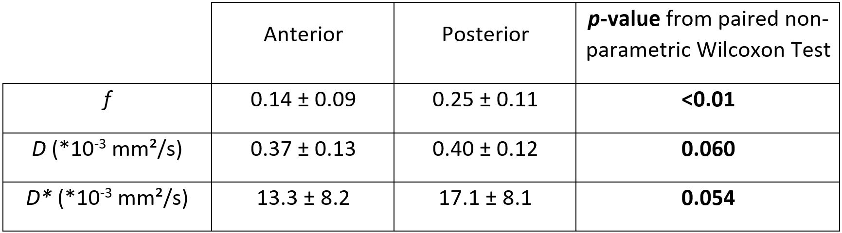
Statistical analysis: Statistical analysis was performed to assess the difference in f, D and D* between posterior and anterior regions. To this purpose, the paired non-parametric signed-rank Wilcoxon test was used with the threshold of p<0.01 for statistical significance.
Results
Figure 1 shows an example of a RESOLVE diffusion-weighted image (b=0 s/mm²) on one volunteer. The posterior and anterior ROIs, drawn on L1 to L5, are illustrated on the image.
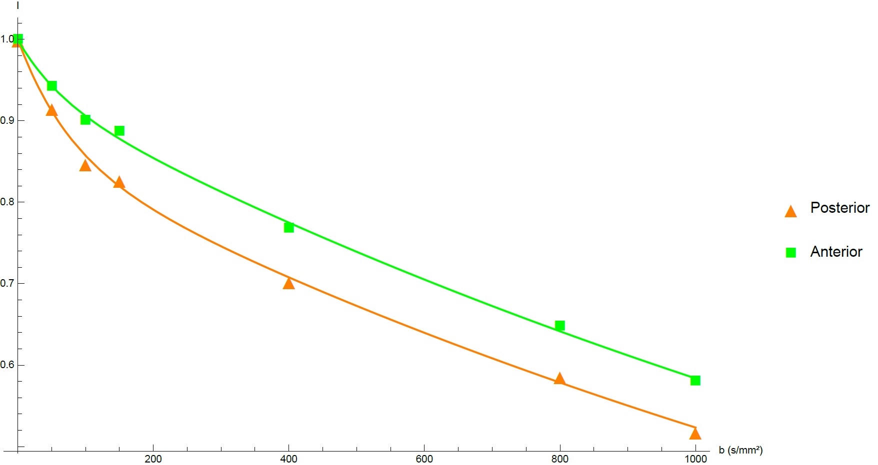
Figure 2 shows an example of IVIM signal decay for posterior and anterior ROIs in the same vertebra. A difference in perfusion between the two IVIM decays is already noticeable by visual inspection. This difference is confirmed by the quantitative analysis of the IVIM parameters, which are summarized in Table1 (posterior ROI vs anterior ROI: f = 0.25 vs 0.14). No significant difference was observed between posterior and anterior ROIs for D and D* [Table1].
Discussion
Only eight studies have been performed to date on IVIM MRI in vertebral bone marrow. All these studies have been conducted in very recent years (2014-2018, references available in the study in reference2). In these studies, the definition of the ROI location was not always well described. In the current study, we have shown a statistical difference in the perfusion fraction between two ROIs (namely, posterior and anterior) of the same vertebra.
Thus, the results shown in the current study indicate that caution has to be taken when drawing ROIs in longitudinal studies. In particular, the location of the ROI has to be 1) well specified and 2) kept constant during along the study. It is also noteworthy that the IVIM protocol, as implemented here with the RESOLVE sequence, provides a very good sensitivity for measurement of f, enabling the detection of regional differences within the same vertebra.
Finally, it should be also noted that, in some studies, the ROIs were chosen to covers the entire vertebral body. Based on the results of the current study, the choice of such ROI would lead to a partial volume effect in the determination of f.
Conclusion
In this study, we have measured regional differences in the IVIM perfusion fraction f between posterior and anterior ROIs. These results highlight the importance of the ROI placement for IVIM measurements in vertebral bone marrow, in particular for longitudinal studies.Acknowledgements
No acknowledgement found.References
1. Karampinos, D. C. et al. Quantitative MRI and spectroscopy of bone marrow: Quantitative MR of Bone Marrow. J. Magn. Reson. Imaging 47, 332–353 (2018).
2. Lasbleiz, J., Le Ster, C., Guillin, R., Saint-Jalmes, H. & Gambarota, G. Measurements of Diffusion and Perfusion in Vertebral Bone Marrow Using Intravoxel Incoherent Motion (IVIM) With Multishot, Readout-Segmented (RESOLVE) Echo-Planar Imaging: IVIM-MRI of Bone Marrow With RESOLVE. J. Magn. Reson. Imaging (2018). doi:10.1002/jmri.26270
Figures