1930
On the Technical Challenges of Diffusion-Weighted MR Spectroscopy for Water ADC Quantification in Human Supraclavicular Fat1Department of Diagnostic and Interventional Radiology, Technical University of Munich, Munich, Germany
Synopsis
The study of brown adipose tissue (BAT) has attracted many researchers in the context of human metabolism. In adult humans, BAT is mostly present in the supraclavicular fossa as a heterogeneous mixture of white and brown adipose tissue. DW MR is a powerful technique to detect microstructural differences which could potentially help to differentiate BAT from surrounding tissue. However, physiological motion in close proximity to the soft adipose tissue in the supraclavicular fossa could lead to signal cancelations and quantification errors. In this work we investigate the feasibility of human BAT DW-MRS for the ADC quantification of the water component in vivo.
Introduction
The study of brown adipose tissue (BAT) has attracted many researchers in the context of human metabolism. The thermogenic nature of BAT in contrast to white adipose tissue (WAT) has been viewed as a potential therapeutic target in obesity treatment1. In adult humans, BAT is mostly present in the supraclavicular fossa as a heterogeneous mixture of WAT and BAT2. Detecting microstructural differences could potentially help to differentiate BAT from the surrounding tissue3 and help to characterize brown adipocytes even further4. Diffusion weighted (DW) magnetic resonance spectroscopy (DW-MRS) is a powerful technique to probe tissue microstructure. However, motion, such as vessel pulsation in the immediate proximity of the BAT can potentially lead to signal cancellations due to intra-voxel dephasing and eventually to an overestimation of the diffusion behavior. In this work we investigate the feasibility and reproducibility of human BAT DW-MRS of the water component in vivo.Methods
A DW bipolar Stimulated Echo Acquisition Mode (DW-STEAM) MRS sequence5 was utilized in combination with ECG cardiac triggering (Fig.1). 8 subjects were scanned on a 3T system (Ingenia Elition, Philips, Best). The sequence parameters were: TE=20ms, TR=3 cardiac cycles, TM=100ms, voxel size (dependent on subject’s anatomy) ~9x9x9mm3, b-values=20,150,220,290,360,430,500s/mm2, 8 averages per polarity. The ECG-based cardiac trigger delay (TD) varied from 500ms to 800ms depending on the subject’s heart rate. First order shimming based on a pencil-beam method (PB volume) was done over the volume. For accurate voxel placement within a potential BAT region, a proton-density fat fraction (PDFF) map was generated from a Multi-Gradient-Echo Dixon technique and the area within the BAT fossa with the lowest PDFF values was selected. To optimize and evaluate the impact of cardiac triggering, DW-MRS at a b-value of 200 s/mm2 and with 16 averages were scanned with varying TD in two subjects beforehand. Separate flow measurements were performed in the axillary artery.
The postprocessing pipeline for apparent diffusion coefficient (ADC) estimation is depicted in Fig.2 and was implemented in MATLAB (MathWorks, Natick, Massachusetts). The processed spectra were fitted to an 8-peak fat model and one water peak. Since fat should not have lost signal due to diffusion at these low b-values, we assumed that the signal loss was due to motion-induced intravoxel dephasing. The water peak area values for the b-values ranging from 150 to 500 s/mm2 were corrected by referencing to the fat peak areas before fitting a mono-exponential function. Two different fitting strategies, an independent fitting (treating every b-value separately and performing an ADC quantification afterwards) and a joined fitting (incorporating the diffusion weighting in the fitting of all spectra) were performed. To test the reproducibility of the measurement a bootstrapping analysis with 30 bootstraps was performed. A random selection of 4 averages out of the acquired 8 averages per polarity was chosen for each bootstrap. Based on the subsequently processed spectra subset a mean value and coefficient of variation (COV) of the ADC for each fitting and subject was obtained.
Results
Fig.3 shows the comparison of the quantitative flow data with the DW-MRS data for different TD in two subjects. For a TD between 400ms and 1100ms the methylene peak area stays at a constant high value and the linewidth of the water peak has a minimum value at around 600ms-700ms. The period of this minimum line width corresponds with a small arterial blood flow velocity. In Fig.4, spectral fitting results for subject #4 and the corresponding diffusion decay curves are illustrated. Within the 8 subjects, three datasets where withdrawn due to either an obvious dislocation of the voxel to neighboring muscle tissue or noisy fat peaks. The table in Fig.5 displays the obtained results for the remaining 5 subjects. Subject #1 had a high PDFF, resulting in a low SNR of water and thus an overall poor fitting quality, as seen in the covariance of the bootstrap analysis. Adding the b-value decay as a constraint for the diffusion decay fitting led to lower CoV values compared to the individual fitting approach.
Discussion & Conclusion
Because of the soft tissue
nature of BAT and the proximity to large vessels, cardiac triggering was found
to improve DW-MRS results (Fig.3) A peak fitting methodology was developed to increase the robustness and precision of water ADC extraction. Fat referencing of water peak areas
resulted in lower water ADC values compared to the analysis without fat
referencing. Joined fitting also reduced water ADC CoV. However, the reported
ADC values and CoV values varied strongly across subjects. Therefore, technical
developments are required to further increase the robustness of water ADC
mapping in the supraclavicular fossa.Acknowledgements
The present work was supported by the European Research Council (grant agreement No 677661, ProFatMRI). This work reflects only the authors view and the EU is not responsible for any use that may be made of the information it contains. The authors would also like to acknowledge research support from Philips Healthcare.
References
[1] Boss O., Farmer S. R. Recruitment of brown adipose tissue as a therapy for obesity-associated diseases. Front. Endocrine. 2012; 3:14
[2] Cypess AM, Haft CR, Laughlin MR, Hu HH. Brown Fat in Humans: Consensus Points and Experimental Guidelines. Cell metabolism. 2014; 20(3):408-415
[3] Verma SK, Nagashima K, Yaligar J, Michael N, Lee SS, Xianfeng T, Gopalan V, Sadananthan SA, Anantharaj R, Velan SS. Differentiating brown and white adipose tissues by high-resolution diffusion NMR spectroscopy. J Lipid Res 2017;58(1):289-298.
[4] Ernande, L.,Stanford, KI., Thoonen, R.,Zhang, H., Clerte, M.,Hirshman, MF.,Goodyear, LJ.,Bloch, KD., Buys, ES.,Scherrer-Crosbie, M., Relationship of brown adipose tissue perfusion and function: a study through β2-adrenoreceptor stimulation. Journal of Applied Physiology. 2016; 120(8): 825-832
[5] Ruschke, S., Kienberger, H., Baum, T., Kooijman, H., Settles, M., Haase, A., Rychlik, M., Rummeny, E. J. and Karampinos, D. C., Diffusion-weighted stimulated echo acquisition mode (DW-STEAM) MR spectroscopy to measure fat unsaturation in regions with low proton-density fat fraction. Magn. Reson. Med. 2016, 75: 32–41
Figures
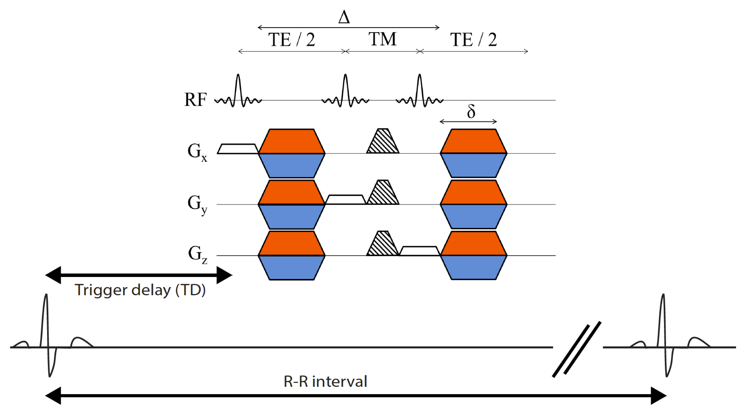
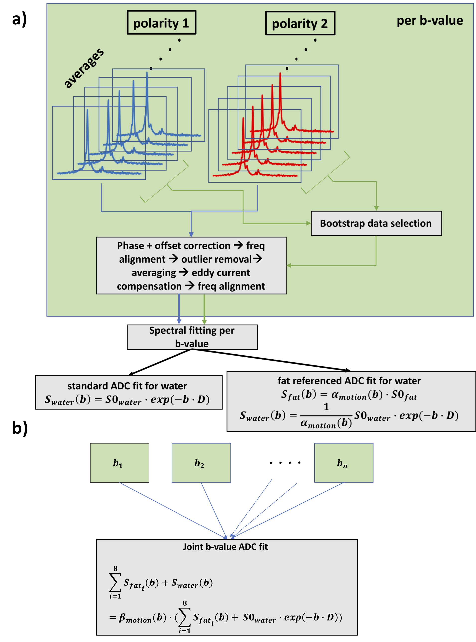
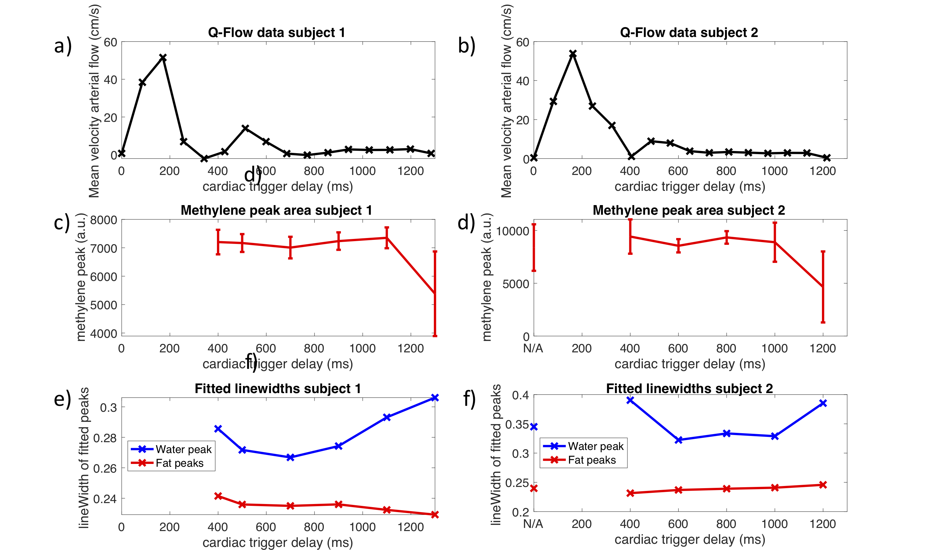
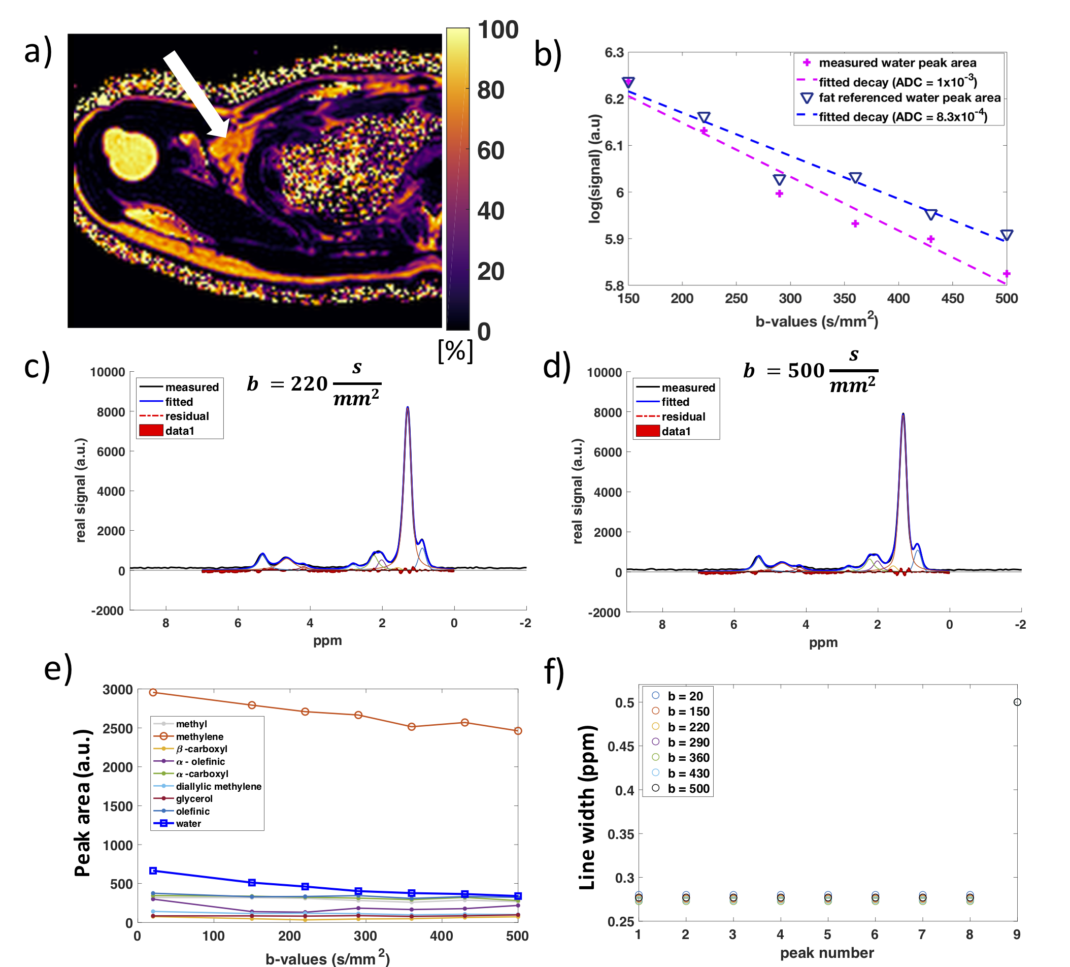
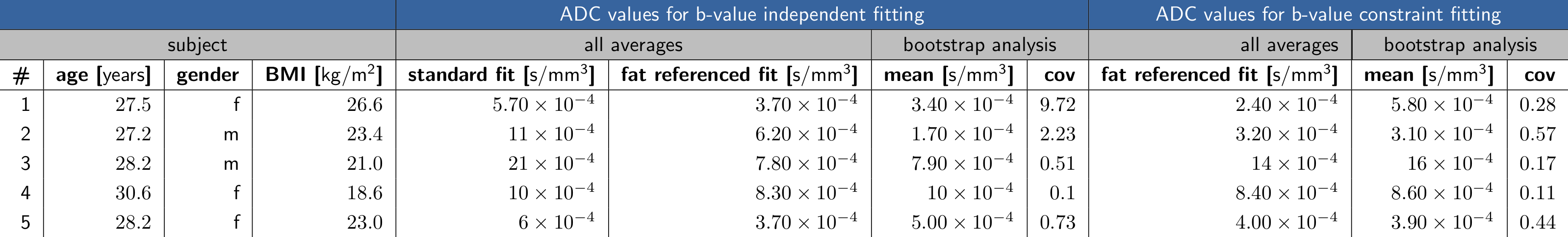
Figure 5: The table shows the ADC estimation results from different fitting approaches. The standard fit leads to overestimated ADC values compared to the fat referenced method. Bootstrap analysis gives information on the reproducibility of each data set, which varies between subjects. Lower CoV values of the bootstrap analysis were found for the joint b-value fitting. In subject 3, the large reduction of the estimated ADC from the standard to the fat referenced method in the case of individual spectral fitting suggests that there was a large amount of intravoxel dephasing due to motion.