1929
White fat browning in a murine model detected by Z-Spectrum Imaging1Radiology, University of Illinois at Chicago, Chicago, IL, United States, 2Bioengineering, University of Illinois at Chicago, Chicago, IL, United States, 3Biophysics and Biophysiology, University of Illinois at Chicago, Chicago, IL, United States, 4Research Resources Center, University of Illinois at Chicago, Chicago, IL, United States
Synopsis
"Browning" of visceral fat is of particular interest to human health due to its association to high risk of metabolic disease and its resistance to conventional browning stimuli. Developing a strategy to stimulate and monitor visceral fat browning could be extremely beneficial in combating epidemic metabolic diseases. Here we report the successful detection of white visceral fat browning in a treated transgenic mouse model by longitudinal Z-Spectrum Imaging. The fat-water fraction measured by ZSI and the fat depots volume were reduced over time compared to the control group. Being noninvasive and with no radiation, the protocol is suitable for longitudinal studies.
Introduction
Brown adipose tissue (BAT) represents one of the brightest hopes in the fight against the obesity epidemic. When activated, this peculiar subtype of fat tissue is able to convert the chemical energy stored in triglycerides and glucose to heat, and has therefore high metabolic relevance on energy expenditure. It has been shown that BAT cell transplants and BAT-overexpressing murine models are more resistant to obesity and other comorbidities [1]. Unfortunately, BAT is not abundant in obese patient and its activation would have limited impact on the overall condition, limiting its application to surgical approaches. The good news is that more and more reports are surfacing about the possibility of converting the widespread white adipose tissue (WAT) to BAT. In fact, “browning” of adipose cells have been observed in subcutaneous and visceral fat [2]. Browning of visceral fat is of particular interest to human health due to its association to high risk of metabolic disease and its resistance to conventional browning stimuli. Developing a strategy to stimulate and monitor visceral fat browning could be extremely beneficial in combating epidemic metabolic diseases. Detection of BAT or browning process have been performed by measuring activity in the form of glucose uptake in 18FDG-PET, or change in MRI biomarkers like T2, T2*, or Fat Water Fraction (FWF). Among the used techniques, Z-Spectrum imaging has advantages due to its merits of being noninvasive, with no radiation and artifacts free [3]. Here we report the successful detection of white adipose tissue browning in a transgenic mouse model by longitudinal Z-Spectrum MRI.Methods
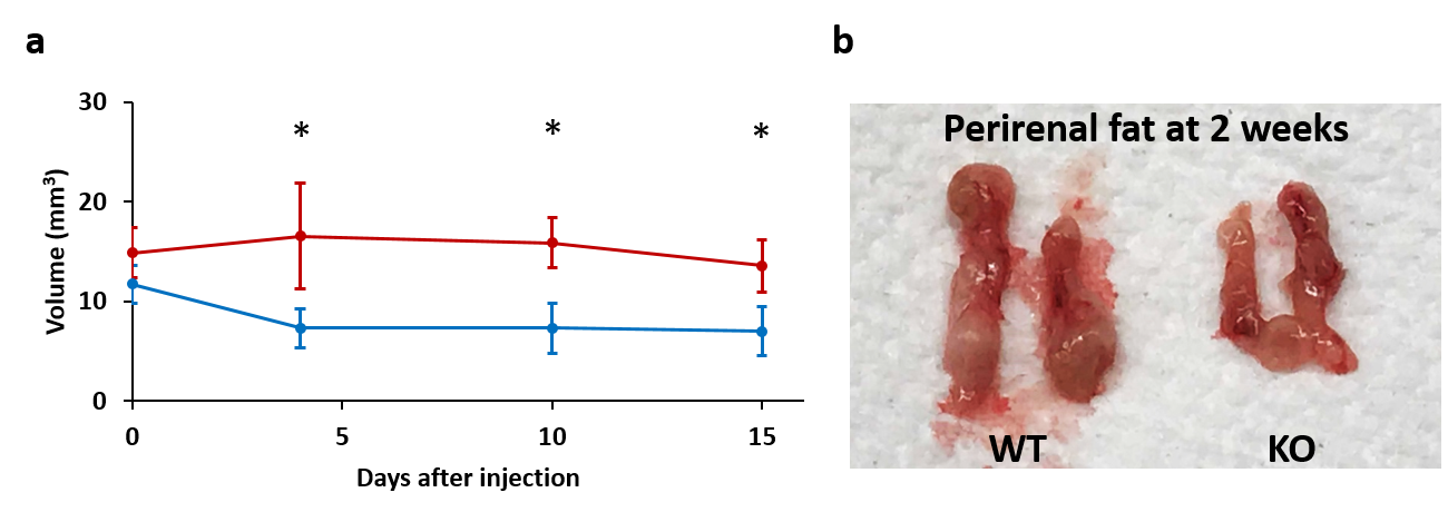
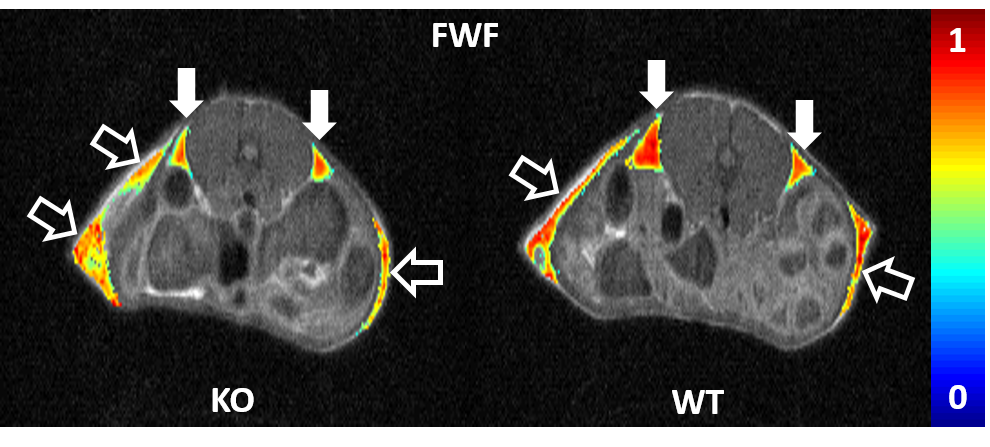
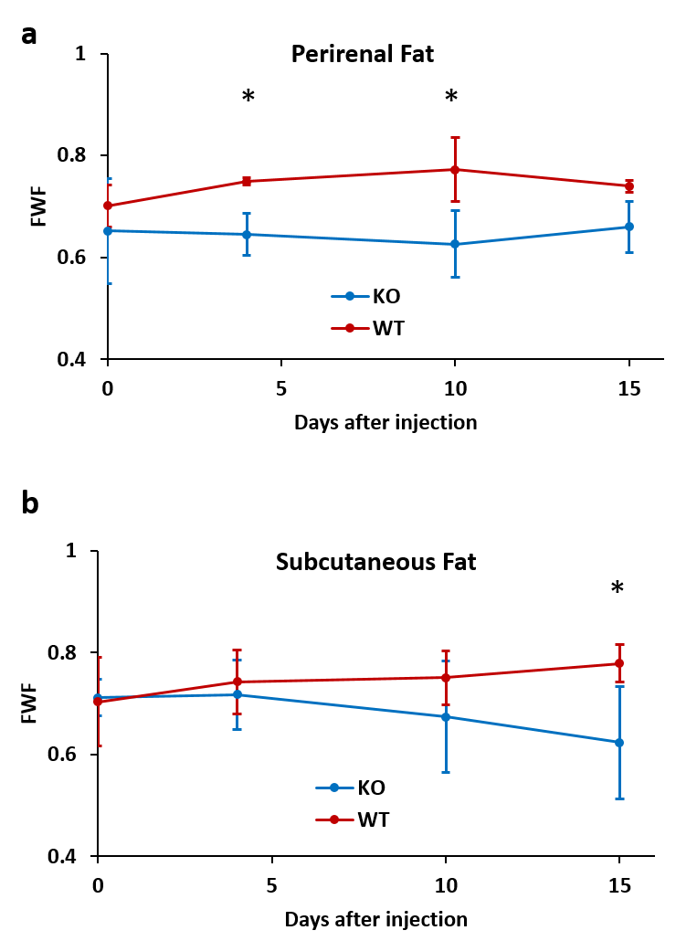
The transgenic murine model was developed by ablation of the transcriptional regulator TRIP-Br2 (TRIP-Br2 KO mice) according to the procedure described in reference [4]. Mice (n=4) were treated IP with 1mg/Kg of β3-AR agonist CL316,243 every day for two weeks. Age-matched wild types (n=3) were similarly treated with saline. Animals underwent MRI at a 9.4T preclinical system before and 4, 10 and 15 days after treatment start. The perirenal white fat depots were identified on scout images and the size of the depots was measured on multislice T2-weighted sequences. Z-Spectrum Imaging was performed with a CEST sequence with a pre-saturation pulse of 0.47µT and 500 msec long, over a range of frequencies spanning ± 5ppm with a 0.1 step size, plus ± 10, +100 ppm for referencing. Saturation was followed by a single slice Fast Spin Echo readout. Z-spectral data were fitted to a multi-Lorentzian model including the direct saturation of water and fat and the magnetization transfer from macromolecules. The fat profile was described by 6 peaks, and the curves’ parameters were loosely constrained to account for variation in lipid composition. FWF was quantified from the fitted amplitudes and colormaps generated for each mouse [3]. Student’s t-test was performed to evaluate significant differences between the groups at each time point.Results and Discussion
Regions of interest analysis showed that the perirenal fat depots decreased in size over time in the KO mice, with volumes shrinking from 11.7±1.9mm3 before treatment to 6.9±2.4mm3 after two weeks. The wild types instead had stable or slightly increased size throughout the study (Fig.1). Fat-water fraction also was found decreased in the drug treated mice compared to the control group (Fig.2). FWF differences between the groups in perirenal fat up to 15% were already detectable after 4 days, and increased to 20% at ten days, but decreased at 2 weeks. Subcutaneous fat showed a sustained decrease in FWF, reaching an average of 25% at the end of the study, but with a higher variability (Fig.3). Both changes in size and FWF are to attribute not only to a higher water content, related to the increased vascular network, but also to a reduced relative lipid content, indicator of brown-like cells sustained activity. Different temporal pattern of variation for perirenal and subcutaneous fat might indicate a hierarchy to the activation mechanism, favoring one fat subtype for the semi-acute response and another for the longer term, as it has been previously suggested [5]. This hypothesis is also supported by the drastic reduction in size of the perirenal depots within the first days, followed by a milder decrease.Conclusion
Z-Spectrum Imaging is proven to be able to detect browning of white adipose tissue. Here we demonstrated the detection of the visceral beige fat of a murine model chronically treated with adrenergic drug. Being noninvasive and carrying virtually no radiation, the protocol is suitable for longitudinal studies, helping to understand the dynamics of chronic activation and assess potential obesity treatment therapies.Acknowledgements
No acknowledgement found.References
1X Liu, Cell Res, 2013; 2A Smorlesi, Obes Rev, 2012; 3AM Scotti, JMRI, 2018; 4CW Liew, Nat Med, 2013; 5J Nedergaard, Cell Metab, 2014.
Figures