1925
Perfusion-Weighted Imaging in Human Kidneys using Hyperpolarized Xenon-1291Medical Faculty Mannheim, Heidelberg University, Computer Assisted Clinical Medicine, Mannheim, Germany, 2Unit of Academic Radiology, University of Sheffield, POLARIS, Sheffield, United Kingdom
Synopsis
Cortical perfusion is an important biomarker that could reflect pathophysiological changes in kidney disease. Novel methods to assess kidney perfusion could be complementary to DCE and ASL techniques. In this work, we demonstrate a proof-of-concept to obtain perfusion-weighted kidney images using time-resolved dissolved hyperpolarized 129Xe MRI. Subtraction of images acquired with different TR showed high contrast within the kidneys that qualitatively match those seen on ASL perfusion maps and indicate higher xenon uptake in the cortex when compared to the medulla. This work demonstrates the potential of hyperpolarized 129Xe as injection free means of assessment of renal perfusion imaging.
Introduction
Cortical perfusion could reflect the pathophysiological processes of kidney diseases1. Arterial spin labelling (ASL) is a promising technique to assess cortical perfusion but it has not been clinically established2. Recently, dissolved hyperpolarized xenon-129 (129Xe) was imaged in the kidneys3. Moreover, dynamic studies using spectroscopy4 and imaging have been presented5, outlining the potential to analyze temporally-dependent physiological processes of the kidney. In this work, we show preliminary perfusion-weighted images of the kidney using dissolved hyperpolarized 129Xe by subtracting dynamic images with different repetition times (TR). The influence of initial bolus arrival times and T1 is also explored.Methods
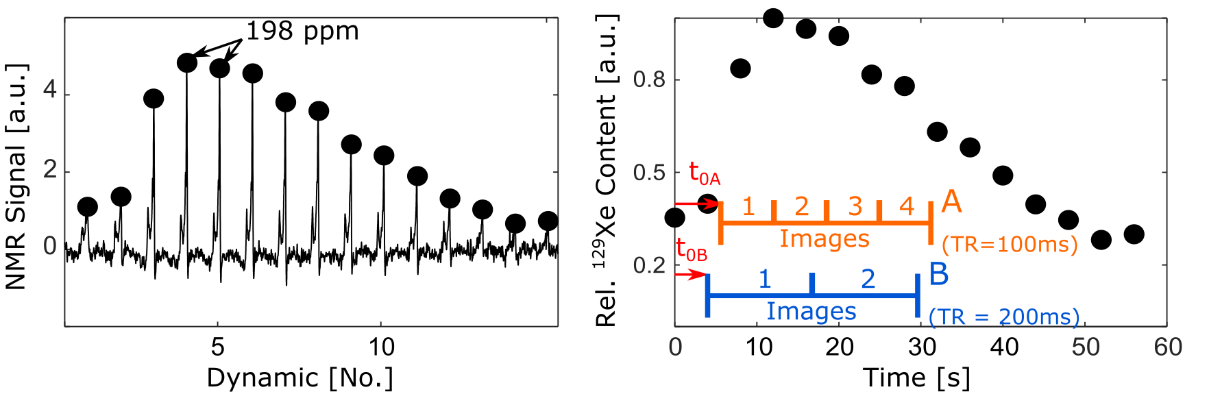
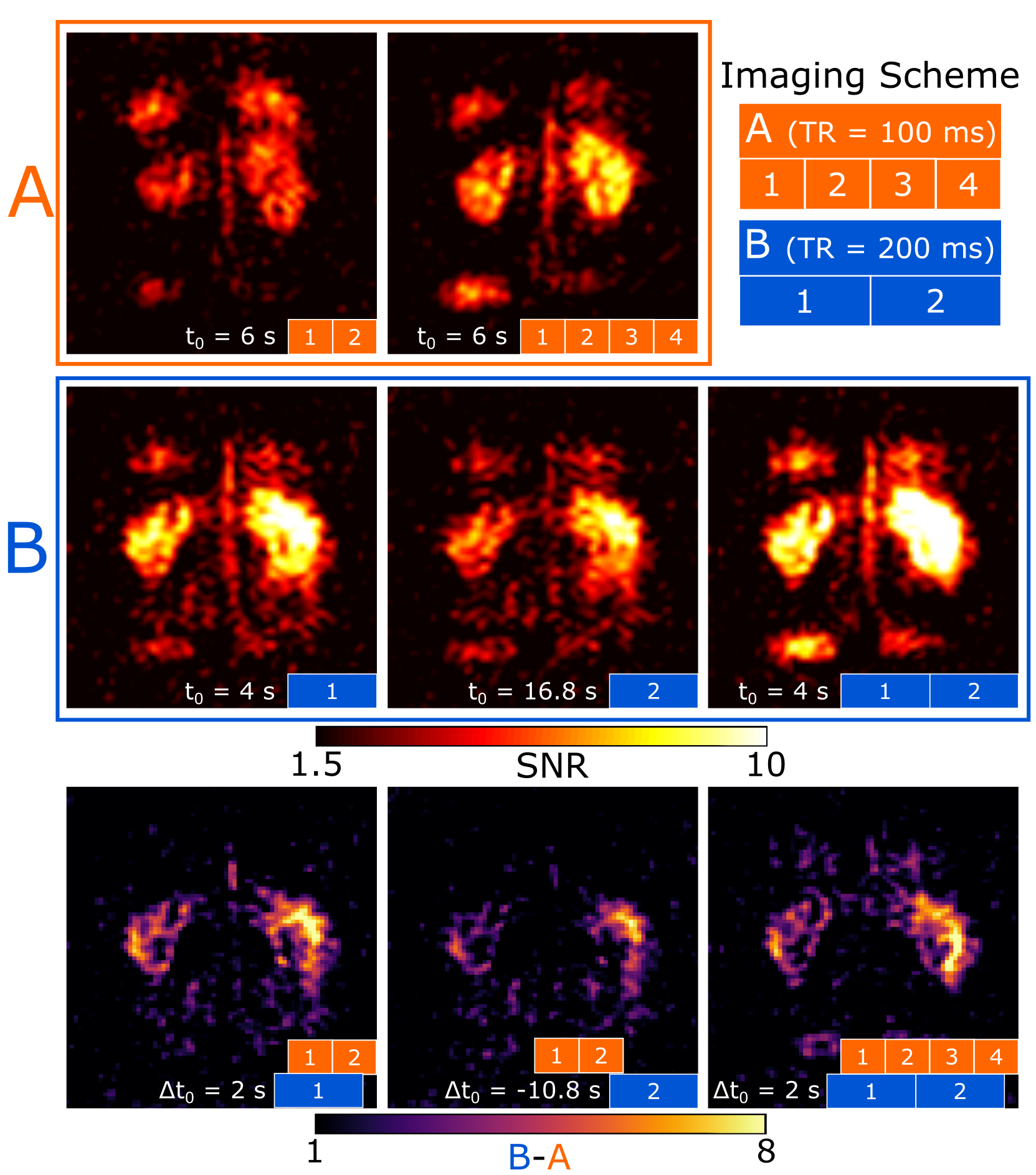
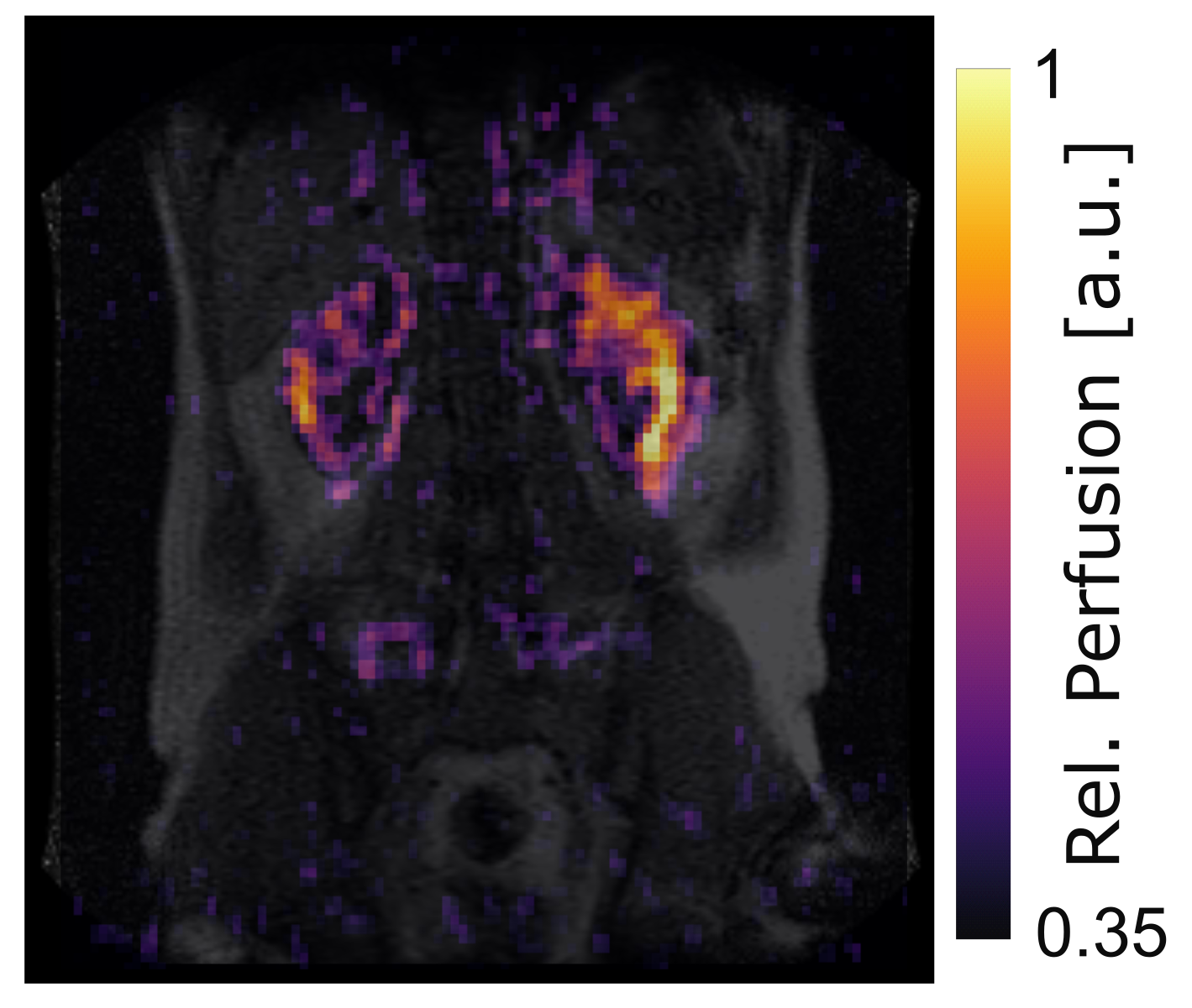
All measurements were performed on a 3T Ingenia MR System (Philips Healthcare, Amsterdam). 129Xe was hyperpolarized in-house using the spin-exchange optical pumping method6. Inhaled doses of xenon constituted 1L of 80% enriched 129Xe gas polarised to ~30%. One healthy volunteer participated in our study. Firstly, dynamic 129Xe pulse-acquire spectroscopy was used to assess the arrival time and relative content of 129Xe in the kidney. Spectroscopy was acquired using a transmit-only-receive-only coil array with sensitivity limited to the kidney5 using the following parameters: TR=4000ms, FA=20°, bandwidth=4096Hz, samples=512, dynamics=15, scan time=56s. Secondly, 129Xe dissolved phase imaging was then performed with a flexible dual Helmholtz quadrature transceiver coil (CMRS, Brookfield, WI) placed around the subjects abdomen, using a spoiled gradient echo sequence (SPGR) with a center frequency of 198ppm from the 129Xe gas reference peak (from the base of the lungs). Two datasets (A and B) were acquired using different repetition times TRA=100ms with four dynamic images and TRB=200ms with two dynamic images, respectively, maintaining an equal total acquisition time. To test the effects of T1 depolarization and initial arrival times of the blood, different delay times (t0A=6s and t0B=4s) were used between full dose inhalation and acquisition start (Fig. 1). Common imaging parameters were: TE=0.80ms, FA=10°, native resolution=8.75x8.75mm², matrix size=64x64, thickness=200mm, total scan time=25.6s. SNR maps from single and selected averaged images were calculated using the standard deviation of the background. Relevant SNR maps from protocol A were subtracted from B (see Fig. 2). 1H scans were acquired with a SPGR sequence (TE/TR/FA=4.6ms/6.2ms/40°) for reference and the 129Xe image was manually superimposed on the corresponding coronal view.Results
Dynamic spectroscopy showed a peak in the kidney at ~198ppm and the maximum NMR signal was observed at 4th dynamic frame (12s) after inhalation of the 129Xe gas (Fig. 1). Imaging yielded higher SNRs for protocol B with a peak (SNR~15) in the left kidney of the averaged two dynamics. No clear image contrast was observed between cortex and medulla in either protocol. Image subtraction (B-A) showed higher SNR differences in the outer (>8) than in the inner kidney and the aorta (<1). The patterns were maintained regardless of Δt0 (Fig. 2). The subtracted 129Xe images conformed with the anatomy in the 1H reference (Fig. 3).Discussion
Dynamic 129Xe spectroscopy helps estimate arrival times and relative 129Xe concentrations using the previously identified plasma/tissue peak4,7-9. Using dynamic imaging, negligible contrast between cortex and medulla was previously shown5 and was also observed here in individual time series A and B. However, by subtracting A from B some distinct contrast was found between the cortex and the medulla corresponding with a higher perfusion expected in the cortex and typical ASL perfusion maps2. This can be explained as follows: dissolved hyperpolarized 129Xe is delivered from the lungs via the heart to the kidneys. Every TR, the RF pulse causes some depolarization of the 129Xe dissolved in the kidneys. Due to a lower RF depolarisation history and the longer T1 of gas in the lungs (~20s10) when compared to the dissolved in blood/kidneys (~8s11), influx of 129Xe with higher polarization will arrive with every heartbeat and replenish the signal. Longer TRs will allow more fresh 129Xe to perfuse the kidney and its distribution will be greater in higher perfused areas i.e. the cortex. T1 depolarization and initial arrival times will influence SNR but the perfusion patterns prevailed despite t0 variations which suggest that TR dominates the observed SNR, and consequently may provide time resolved perfusion-weighting of the 129Xe signal in the kidney. In further work, modeling the effect of the FA and B1 homogeneity, TR and T1 on 129Xe spatio-temporal signal modulation combined with 1H ASL information should shed further light on the perfusion-weighting imaging we observe.Conclusion
Hyperpolarized dissolved 129Xe indicates sensitivity to cortical perfusion based on images acquired with different TRs. In the future, further development of this principle should be a valuable addition to the existing tools to evaluate kidney diseases.Acknowledgements
This work was funded in part by the MRC MR/M008894/1 and EU. This article/publication is based upon work from COST Action CA16103, supported by COST (European Cooperation in Science and Technology). www.cost.eu.References
1. Li LP, Tan H, Thacker JM, et al. Evaluation of Renal Blood Flow in Chronic Kidney Disease Using Arterial Spin Labeling Perfusion Magnetic Resonance Imaging. Kidney Int Rep 2017;2(1):36-43.
2. Odudu A, Nery F, Harteveld AA, et al. Arterial spin labelling MRI to measure renal perfusion: a systematic review and statement paper. Nephrol Dial Transplant 2018;33(suppl_2):ii15-ii21.
3. Mugler III JP, Miller GW, Meyer CH, et al. Imaging of dissolved-phase hyperpolarized xenon-129 in human kidneys. In Proceedings of the 23th Annual Meeting of ISMRM, Toronto, Canada 2015;23:848.
4. Miller GW, Cates Jr. GD, Keder D, et al. Dynamic Spectroscopy of Dissolved-Phase Xenon-129 in the Human Kidney. In Proceedings of the 25th Annual Meeting of ISMRM, Honolulu, USA 2017;25:1182.
5. Chacon-Caldera J, Maunder AM, Rao M, et al. Dynamic MRI of Hyperpolarized Xenon-129 Uptake in the Human Kidney Using a Dedicated Transmission-Only-Reception-Only Array at 3 Tesla. In Proceedings of the 26th Annual Meeting of ISMRM, Paris, France 2018;26:4470.
6. Norquay G, Collier GJ, Rao M, et al. 129Xe-Rb Spin-Exchange Optical Pumping with High Photon Efficiency. Phys Rev Lett 2018;121(15), 153201.
7. Antonacci MA, Zhang L, Burant A, et al. Simple and robust referencing system enables identification of dissolved-phase xenon spectral frequencies. Magn Reson Med 2018;80(2):431-441.
8. Swanson SD, Rosen MS, Coulter KP, et al. Distribution and dynamics of laser-polarized (129)Xe magnetization in vivo. Magn Reson Med 1999;42(6):1137-45
9. Rao M, Stewart NJ, Norquay G, et al. High resolution spectroscopy and chemical shift imaging of hyperpolarized (129) Xe dissolved in the human brain in vivo at 1.5 tesla. Magn Reson Med 2016;75(6):2227-34
10. Kruger SJ, Nagle SK, Couch MJ, et al. Functional imaging of the lungs with gas agents. J Magn Reson Imaging. 2016;43(2), 295-315.
11. Norquay G, Leung G, Stewart NJ, et al. Relaxation and exchange dynamics of hyperpolarized 129Xe in human blood. Magn Reson Med. 2015;74(2):303–311.
Figures