1923
Whole Body Quadrature Birdcage RF Coil for 23Na MRI of Small Rodents at 9.4T and Its Application in Sodium Imaging of the Eye and Kidneys1Berlin Ultrahigh Field Facility (B.U.F.F.), Max Delbrück Center for Molecular Medicine in the Helmholtz Association, Berlin, Germany, 2MRI.TOOLS GmbH, Berlin, Germany, 3Berlin Ultrahigh Field Facility (B.U.F.F.), Max Delbrück Center for Molecular Medicine in the Helmholtz, Berlin, Germany, 4Institute of Vegetative Physiology, Charité - University Medicine Berlin, Berlin, Germany
Synopsis
Sodium ions play a major role in the physiology of the human organism. The kidneys and eyes both have a high sodium concentration, and we suggest that probing tissue sodium concentration in these organs using 23Na MRI can add a very useful dimension to our understanding of renal and ocular disorders. Changes in the sodium concentration might be indicative of early pathophysiological changes in such diseases. Therefore we present a quadrature birdcage coil tailored for sodium imaging of small rodents at 9.4T, along with the RF coil design, EMF simulations and in vivo 23Na MRI of the eye and kidney.
Introduction
Sodium ions (Na+) play an important role in the physiology of the human organism, with the sodium/potassium pumps being essential for the maintenance of homeostasis1. High Na+ concentrations are found in the renal medulla and in certain compartments of the eye. In the kidney, tubular water resorption is driven by the osmotic gradient between the tubuli and the interstitium that largely relies on active transport of sodium2. Active transport of Na+ also plays an important role in the human eye and its compartments, including the formation of the aqueous humor and the removal of the water and lactic acid from the retina3. Dysfunction in these processes could lead to changes in the sodium concentration and might be symptomatic of early pathophysiological changes in renal and ocular diseases4,5. 23Na MRI could add a very useful dimension to our understanding of renal, and ocular disorder but also for detailing differences between sodium deposition in muscle and skin in cardiac and environmental metabolic diseases. Recognizing the potential of whole body 23Na MRI in animal models this work describes the design, construction and performance of a large volume 23Na quadrature birdcage radiofrequency (RF) coil tailored for rodents at 9.4T. The applicability of the proposed whole body RF coil is demonstrated for in vivo 23Na MRI of the rat eye and kidney.Methods
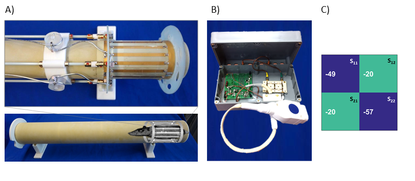
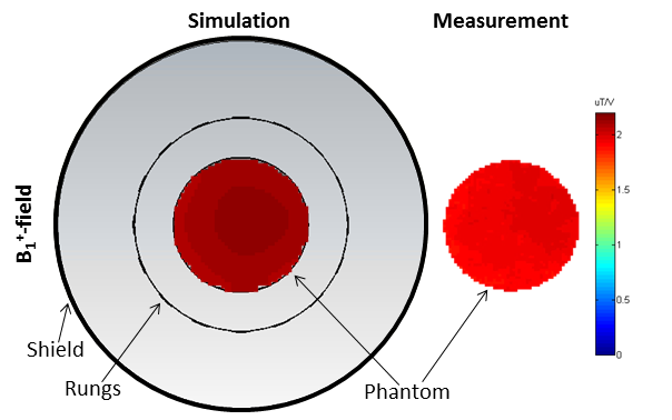
The inner diameter of 62mm of the proposed low pass birdcage was given by the size of a typical loading (small rat) and the size of the MR scanner bore and contains 16 rungs (Figure 1A). Birdcage Builder6 was used in order to estimate the initial values of distributed capacitors. EMF simulations were carried out using CST Studio Suite 2016 and included: a rat-shaped 3D model (σ=0.91S/m, ε=65) (Figure 1B); and a cylindrical phantom (σ=0.72S/m, ε=63). RF coil bench measurements were performed on a saline phantom (V=200mL, [NaCl]=600mM, σ=0.72S/m, ε=63) using a network analyzer. To drive the birdcage (Figure 2A) in quadrature mode, an additional T/R switch and hybrid combiner was designed and built (Figure 2B). MR experiments were conducted on a 9.4T animal scanner (Biospec, Bruker, Germany). B1+-field maps were acquired using double-angle-method7. For reference image a 1H birdcage coil with same geometry as the 23Na birdcage was used. The change to the quadrature driven 23Na birdcage was done without moving the animal. 23Na eye imaging required placing the rat’s head in the center of the birdcage coil, which resulted in a change in the coupling between the sample and the end ring of the coil. This change was compensated by adding a loading phantom.Results
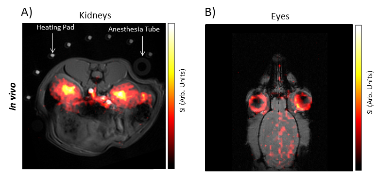
Figure 2C represents the S-parameter values measured on a homogeneous phantom. The reflection coefficients (S11, S22) were measured to be -49dB or better and the transmission coefficient (S12, S21) was -20dB. The transmit field (B1+) simulation result was validated in a MR experiment. Figure 3 shows the simulated RF coil design in axial orientation and the corresponding B1+-map for the virtual phantom and the measured B1+-map for the cylindrical phantom for the same ROI. The B1+-profile over the cross-section of the phantom ranged from 1.8-2.0µT/V for the measurement and 2.0-2.3µT/V for the simulation. Considering the uncertainty of losses in the RF-coil (copper, solder and capacitors), the magnitude of the measured and simulated B1+-fields are in good agreement. Examples of in vivo 23Na MRI of the rat kidneys and eyes are shown in Figure 4A-B using an overlay of 1H and 23Na MR images. For renal and ocular 23Na MRI an in-plane spatial resolution as good as 780µm was achieved. This resolution and sensitivity facilitates the visualization of the renal corticomedullary sodium gradient. Our results also show that the sodium content in the lens is distinguishable from the sodium content in the aqueous and vitreous humor.Discussion and Conclusion
This work reports on the design, construction and feasibility of a whole body 23Na quadrature birdcage RF coil tailored for rats at 9.4T. Equipped with this 23Na RF technology, we will commence animal studies to elucidate the intertwined roles of renal tissue hypoperfusion, hypoxia, energy metabolism and sodium metabolism en route to developing novel treatment options and effective prophylactic regimens for acute kidney injury and chronic kidney disease. Likewise, the proposed 23Na MRI approach will allow the assessment of the sodium content in the eye and its compartments in vivo as part of our translational research in humans and in animal models8. The broad roles of this sodium in processes related to eye physiology suggest a wide range of questions for in vivo ophthalmological investigations afforded by 23Na MRI.Acknowledgements
This work was supported in part by the Bundesministerium für Bildung und Forschung (BMBF, German Federal Ministry for Education and Research; grants VIP+ 03P00081, VIP+ 03P00082).References
1. Murphy, E. and D.A. Eisner, Regulation of intracellular and mitochondrial sodium in health and disease. Circ Res, 2009. 104(3): p. 292-303.
2. Douglas C. Eaton, J.P.P., Vander's Renal Physiology. 7th ed2009: Mc Graw Hill LANGE.
3. Kaufman PL, A.F., Levin LA, Alm A, Adler's Physiology of the Eye 2011: Elsevier Health Sciences.
4. Garner, W.H., et al., Sodium-23 magnetic resonance imaging of the eye and lens. Proc Natl Acad Sci U S A, 1986. 83(6): p. 1901-5.
5. Maril, N., et al., Detection of evolving acute tubular necrosis with renal 23Na MRI: studies in rats. Kidney Int, 2006. 69(4): p. 765-8.
6. Chin, C.L., et al., BirdcageBuilder: Design of Specified-Geometry Birdcage Coils with Desired Current Pattern and Resonant Frequency. Concepts Magn Reson, 2002. 15(2): p. 156-163.
7. Akoka, S., et al., Radiofrequency map of an NMR coil by imaging. Magn Reson Imaging, 1993. 11(3): p. 437-41.
8. Wenz, D., et al., Millimeter spatial resolution in vivo sodium MRI of the human eye at 7 T using a dedicated radiofrequency transceiver array. Magn Reson Med, 2018. 80(2): p. 672-684.
Figures