1921
The Effect of Water Loading on Renal Diffusion Parameters with Triexponential Analysis1Department of Radiological Technology, Kanazawa University Hospital, Kanazawa, Japan, 2Faculty of Health Sciences, Institute of Medical, Pharmaceutical and Health Sciences, Kanazawa University, Kanazawa, Japan, 3Department of Radiology, Kanazawa University Hospital, Kanazawa, Japan
Synopsis
To assess the response of water loading on diffusion parameters in the renal tissue, we evaluated renal diffusion coefficients with tri- and biexponential analyses before and after water loading. Our results showed that slow-restricted diffusion coefficient with triexponential analysis in the renal medulla was significantly increased after water loading, whereas none of the diffusion parameters with biexponential analysis significantly changed. Diffusion analysis with triexponential function could enable us to obtain more detailed information on renal function.
INTRODUCTION
Intravoxel incoherent motion (IVIM) analysis with biexponential function provides both perfusion and diffusion information and has the potential to assess renal dysfunction.1 A previous study has shown the presence of other two diffusion components in the renal tissue, i.e., fast-free and slow-restricted diffusion, in addition to the perfusion component.2 Thus, diffusion analysis with triexponential function (assuming these three components: perfusion-related diffusion, fast-free diffusion, and slow-restricted diffusion) may enable noninvasive gathering of more detailed information on renal perfusion and diffusion.2-4 Moreover, water loading as a functional challenge for the kidney can be performed to detect premature renal dysfunction.5 However, the effect of water loading on triexponential diffusion analysis has yet to be clarified. Therefore, in this study, to assess the response of water loading on diffusion parameters in the renal tissue, we evaluated these diffusion coefficients with tri- and biexponential analyses at baseline and after water loading.METHODS
On a 3.0-T MRI, coronal diffusion-weighted images of the kidney were acquired in seven healthy volunteers (all men; mean age, 23.3±1.7 years) using single-shot diffusion echo-planar imaging with respiratory triggering. The imaging parameters were: repetition time, one respiration cycle; echo time, shortest; acquisition matrix, 96 × 124; b-values, 0, 10, 30, 50, 100, 200, 400, 600, 800, 1000, and 1200 s/mm2; separate diffusion measures in three orthogonal directions; field of view, 300 mm; slice thickness, 7 mm; number of signals averaged, 2; half-scan factor, 0.633; and parallel imaging factor, 2. All subjects were asked to fast for at least 5 hours before the experiment. Baseline data were acquired prior to water loading. They were then asked to drink 15 mL of water per kilogram of body weight to induce water diuresis and rescanned 1 hour after the start of water loading. In this study, right kidney images were analyzed because left kidney images were degraded due to cardiac-induced motion artifact. We determined mean signal intensities in the renal cortex and medulla at each b-value. Next, perfusion-related, fast-free, and slow-restricted diffusion coefficients (Dp, Df, and Ds, respectively) were calculated using triexponential fitting. Note that we assigned Df to the literature value of the diffusion coefficient of free water at 37 degrees centigrade (3.0 × 10⁻³ mm2/s) in the same manner as a previous study.4 In addition, we also calculated perfusion-related diffusion coefficient (D*) and perfusion-independent diffusion coefficient (D) using biexponential fitting. These diffusion parameters were compared at baseline and after water loading.RESULTS AND DISCUSSION
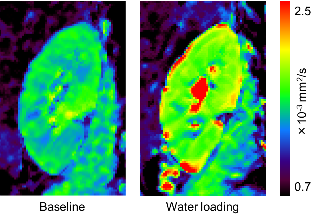
Tables 1 and 2 show diffusion parameters with tri- and biexponential analyses in the renal cortex and medulla, respectively, at baseline and after water loading. Representative Ds images of the right kidney at baseline and after water loading are presented in Figure 1. Ds in the medulla was significantly increased after water loading, whereas medullary diffusion parameters with biexponential analysis did not significantly change. These findings may be explained by the fact that water loading induces functional changes in the medulla, including less restriction of water diffusion from tubule dilation and increase in tubular flow from increased urine production.6 These results also indicate that triexponential analysis could extract the functional change in the medulla better than biexponential analysis. In contrast, none of the diffusion parameters in the cortex was significantly changed after water loading.CONCLUSION
Slow-restricted diffusion in the renal medulla is the most sensitive to functional changes 1 hour after water loading. Diffusion analysis with triexponential function could enable us to obtain more detailed information on renal function.Acknowledgements
No acknowledgement found.References
1. Bane
O, et al. J Magn Reson Imaging. 2016;
44: 317-326.
2.
Baalen S, et al. J Magn Reson Imaging.
2017; 46: 228-239.
3. Hayashi T, et al. J Magn Reson Imaging. 2013; 38: 148-153.
4.
Ohno N, et al. J Magn Reson Imaging.
2016; 43: 818-823.
5.
Kalis IM, et al. Magn Reson Med.
2017; 77: 1573-1582.
6.
Sigmund EE, et al. Radiology. 2012;
263: 758-769.
Figures