1919
Use of Intravoxel incoherent motion imaging in assessment of different degrees of renal lesions: alterations in diffusion and perfusion in animal study with renal lesions1Peking University, Beijing, China, 2Shanghai Jiao Tong University, Shanghai, China, 3Peking University First Hospital, Beijing, China
Synopsis
IVIM DWI (intravoxel incoherent motion diffusion-weighted image) is a noninvasive imaging technology capable of simultaneously detecting diffusion and perfusion characteristics of renal tissue . This study investigates the feasibility of IVIM DWI in evaluating different degree of renal lesions. IVIM DWI was performed on sixteen rabbits with different degree of renal lesions, which were confirmed by the corresponding pathological results. A noticeable change of renal diffusion and perfusion can be clearly observed in different degrees of renal lesions. There exists significant difference in f values among four groups with different severity in renal lesions.
Introduction
Purpose
To investigate the feasibility of using IVIM DWI to distinguish the different degree of renal lesions in renal lesions animals.Methods
Animal Model:
All procedures used in this study were approved by our local Research Ethics Committee. Sixteen New Zealand rabbits with body weights of 2.0-3.5 kg were included in the study. To establish renal lesions model, we injected 0.8 mg microspheres (acryl beads, 40 to 120μm in diameter) into the right renal artery under anesthesia condition. MRI scan was performed to evaluate different degree of renal lesions. Pathological images of H&E staining were obtained four weeks later.
MR Imaging:
IVIM DWI was performed on rabbits (n = 16) using a 3.0 T MRI scanner (Achieva, Philips Medical Systems). During the MRI experiments, anesthesia was induced with isoflurane (1.0 L/min) delivered by a facemask. DWI was performed using a single-shot spin-echo echo-planar (EPI) sequence (coronal, TR = 3000 ms, TE = 65 ms, acquired matrix = 98 × 80, FOV = 150 × 150 mm2, slice thickness = 5 mm, gap = 1.0 mm, SNESE factor = 2). The following b-values were used for acquisition: 0, 20, 40, 60, 80, 100, 150, 200, 300, 400, 500, 600, 700, 800, 900 and 1000 sec/mm2. Respiratory triggering was applied to make the images motion insensitive. According to the IVIM model, the DWI signal can be related as follows:
Sb/S0 = (1-f)e-bD+fe-bD*
where S0 is the signal intensity at b = 0, Sb is the signal intensity at nonzero b value, D is the true diffusion coefficient, D* is the pseudodiffusion coefficient, and f is the fraction of microvascular volume. All the parameters were estimated using the least squares fitting algorithm. The changes in the diffusion (D) and perfusion parameters (D* and f) were analyzed. Regions of interest (ROIs) were placed in each right kidney by an experienced abdominal radiologist. Student’s t-test was used to compare the parameters between the two groups.
Results
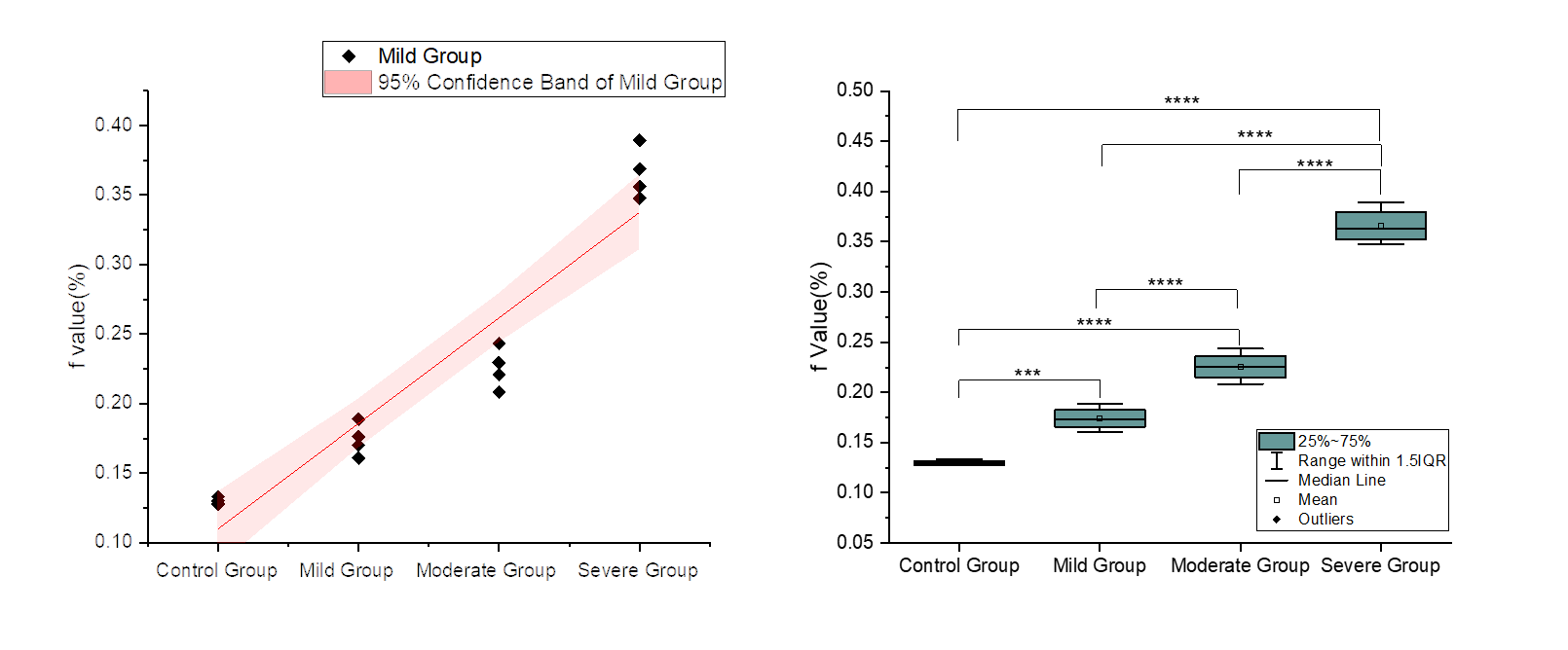
Fig 1 shows representative DWI images acquired different degrees of renal lesions. Fig 2 shows the renal parametric maps of IVIM-metrics (f, D and D* maps) of four representative rabbits with different degrees of renal lesions. A noticeable change of renal diffusion and perfusion can be seen in different degrees of renal lesions (Fig 2, red arrows), which is consistent with previous reports [3]. Pathological results confirmed the renal lesions with findings of wrinkled features with dilated change of Bowman’s capsule (Fig 3). For the four representative rabbits with different degrees of renal lesions D, f and D* have different patterns of change. The D values of renal lesions were significantly higher in mild group (1.7 ± 0.1 × 10⁻³ mm2/s, p < 0.01) than control group (1.23 ± 0.21 × 10⁻³ mm2/s). Besides, the D values were lower in moderate group (1.07 ± 0.1 × 10⁻³ mm2/s, p < 0.01) than mild group (1.7 ± 0.1 × 10⁻³ mm2/s). The D values were significantly lower in severe group (0.76 ± 0.3 × 10⁻³ mm2/s, p < 0.05) than control group (1.23 ± 0.21 × 10⁻³ mm2/s). The f values of renal lesions were significantly higher in in mild group (0.174 ± 0.011, p < 0.001), moderate group (0.225 ± 0.014, p < 0.001), and severe group (0.365 ± 0.017, p < 0.001) than control group (0.129 ± 0.002). Significant differences were found in f among groups with different severities of renal lesions (normal, mild, moderate, and severe, p < 0.001). Besides, the f values of the renal lesions were significantly correlated with pathological results (Fig 4). The D* values of renal lesions were significantly lower in mild group (6.2 ± 2.5 × 10⁻³ mm2/s, p < 0.05) than control group (9.5 ± 1.4 × 10⁻³ mm2/s).Conclusion:
Acknowledgements
No acknowledgement found.References
1. Bellomo R, Ronco C, Kellum J A, et al. Acute renal failure–definition, outcome measures, animal models, fluid therapy and information technology needs: the Second International Consensus Conference of the Acute Dialysis Quality Initiative (ADQI) Group[J]. Critical care, 2004, 8(4): R204.
2. Lameire N, Van Biesen W, Vanholder R. The changing epidemiology of acute renal failure[J]. Nature Reviews Nephrology, 2006, 2(7): 364.
3. Notohamiprodjo M, Chandarana H, Mikheev A, et al. Combined intravoxel incoherent motion and diffusion tensor imaging of renal diffusion and flow anisotropy. Magn Reson Med 2015, 73:1526–1532
Figures