1914
High resolution multi-echo time-of-flight angiography of the kidney at 3 and 7T1SPMIC Department of Physics, University of Nottingham, Nottingham, United Kingdom
Synopsis
We have used a multi-echo time-of-flight angiography sequence to provide increased signal-to-noise ratio, by summing the echoes, to image the kidney at 0.75 mm in-plane resolution and visualize the small distal vessels without the use of contrast agent. Clear images could be obtained at 7T, but results at 7T were not consistently good across all subjects due to limitations in RF power and shimming.
Introduction
A large component of chronic kidney disease (CKD) in the elderly is thought to be “vascular” affecting smaller arteries and arterioles rather than the main renal artery, current imaging methods lack the sensitivity to detect such changes. Multi-echo time-of-flight (TOF) angiography has been proposed to increase sensitivity1.
Aim: To develop multi-echo TOF for high sensitivity angiography at 3 and 7T2 to image more distal vessels at the cortico-medullary border at high spatial resolution.
Methods
TOF angiography was acquired in four subjects across 3 scanners. Data
was collected on a 7T Philips Achieva scanner with an MRCoils 8chTx/32chRx
dipole array body coil, a 3T Philips Ingenia (digital Rx) scanner with a 32ch
torso receive coil, and a 3T Philips Achieva scanner with a 32ch torso receive
coil. A 2D standard multiecho fast field echo sequence was used: TR= 30-39ms, 4
gradient echoes with flyback, TE/ΔTE= 5.3-7.9/7.0 – 8.9 ms,
resolution 0.75 x 0.75 x 2.0 mm, 6 slices, SENSE 2.5. The target flip angle was
50o at 7T (optimized in pilot measurements and not always
achievable), and 50o at 3T. At 7T B1+ shimming was
applied using a phase nulling method in regions of interest encompassing the
right kidney. A B1+ map (DREAM method) was acquired after the shimming
was performed to determine the achievable flip angle. At 7T, B0
shimming was performed using Philips volume shimtool. Maximum Intensity Projections
(MIPs) were created from the first echo, and from the sum of all four echoes.
Results
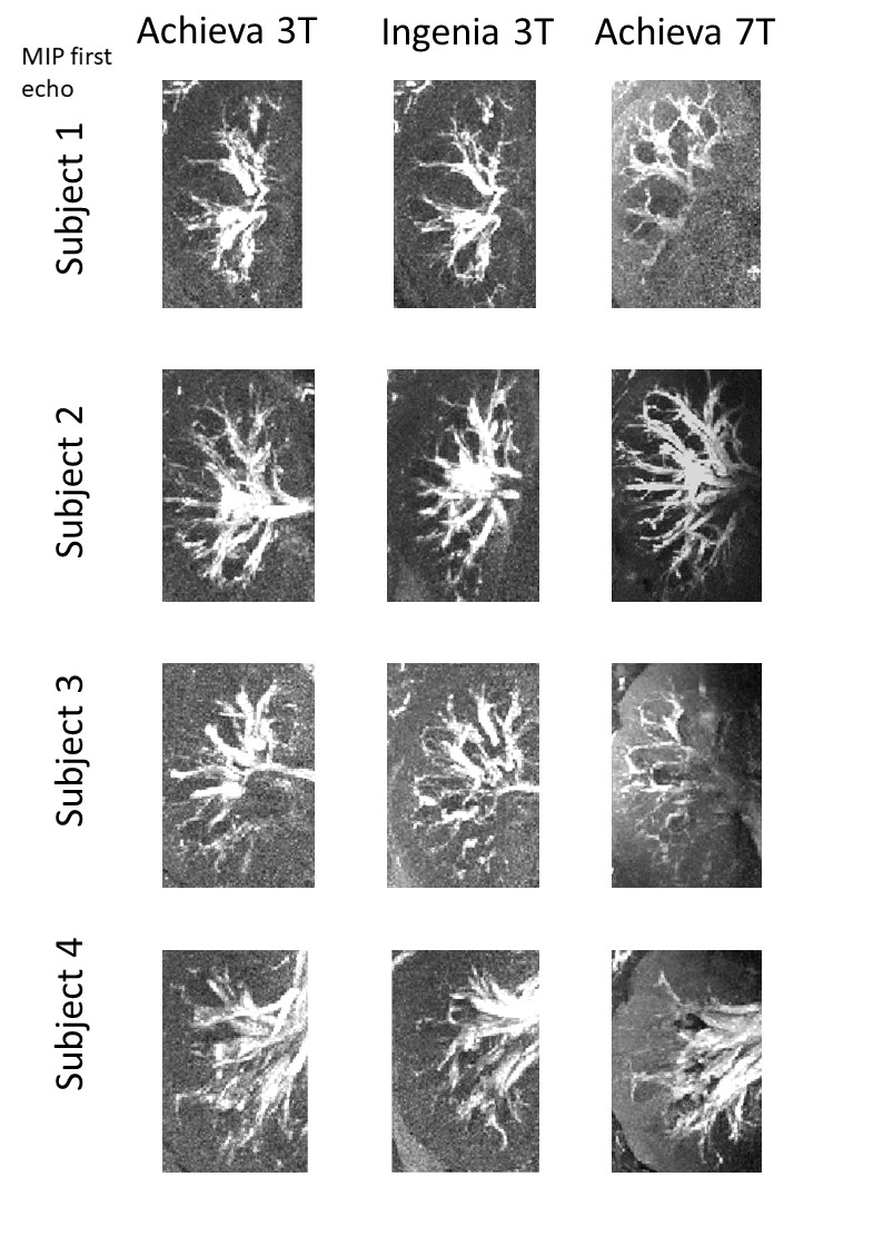
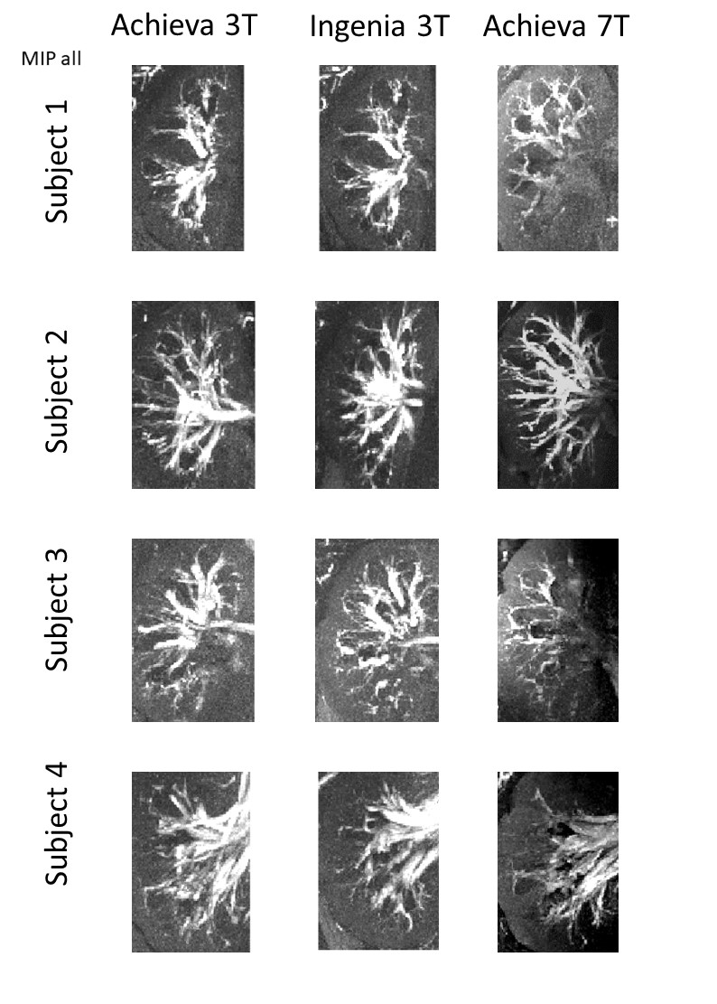
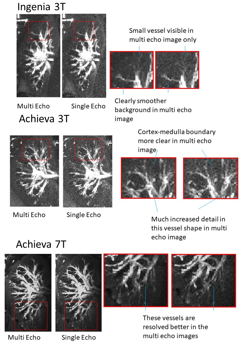
Figure 1 shows example angiogram MIPs from the first echo time only at 3T and 7T. Figure 2 shows corresponding angiogram MIPs from summing across all four echoes. This can be seen to increase vessel-to-background contrast for improved visualization of smaller vessels at the cortico-medullary border. It can be seen that when it worked well (eg Subjects 1 and 2), the 7T data gave the best performance particularly in the cortex, although the 3T data was better at consistently providing high quality data across the whole body of the kidney. Figure 3 highlights the improved visualization of small vessels by summing multiple echoes.
Discussion
Multi-echo TOF provides the increased
contrast-to-noise to show the delineation of small intrarenal arteries at high
spatial resolution (0.75 x 0.75 x 2 mm3). It is a challenge to
acquire high spatial resolution images in a breath-hold. In this work, scan
time was reduced by increasing the SENSE factor and compensating for reduced
signal-to-noise by summing the multi-echo images. At 7T the longer T1 relaxation
time can provide CNR particularly in small peripheral vessels, as seen in
Subjects 1 and 2. However the 7T results can be unpredictable, particularly
since there is often an RF drop out in the middle of the body which can prevent
excitation of the inflowing blood. Tailored RF excitation might be useful in
this regard.
Conclusion
Multi-echo TOF allows improved visualisation of the smaller arteries and arterioles within the kidney. 7T can produce high quality images of the small peripheral vessels but the results can be unstable with current shimming methods and available RF power.
Acknowledgements
Medical Research Council (MRC)
Engineering and Physical Sciences Research Council (EPSRC)
Oxford-Nottingham Biomedical Imaging Doctoral Training Centre (ONBI DTC)
References
1.
Dumoulin CL, Souza SP, Feng H. Multiecho
magnetic resonance angiography. Magn Reson Med. 1987 Jul;5(1):47-57.
2. Grochowski C, Staśkiewicz G. Ultra high field
TOF-MRA: A method to visualize small cerebral vessels. 7T TOF-MRA sequence
parameters on different MRI scanners - Literature review. Neurol Neurochir Pol.
2017 Sep - Oct;51(5):411-418.
Figures