1910
Feasibility of DKI in distinguishing stage IA and IB of endometrial carcinoma1radiology, the first Affiliated Hospital of Dalian Medical University, dalian, China, 2the First Affiliated Hospital of Dalian Medical University, dalian, China, 3GE Healthcare, Beijing, China
Synopsis
Preoperative information about depth of myometrial invasion is therefore essential in tailoring the surgical approach for patients in stage I. Consequently, we investigate the feasibility of diffusion-kurtosis imaging(DKI) in distinguishing the stage IA and IB of EC.
Introduction
Endometrial carcinoma (EC) is one of the common gynecologic malignancies. Tumors confined to the endometrium and those invading the superficial myometrium are designated as stage IA, and tumors invading the deep myometrium are designated as stage IB [1]. Depth of myometrial invasion is the most important morphologic prognostic factor [1]. The incidence of lymph node metastases increases from 3% with superficial myometrial invasion to 46% with deep myometrial invasion. Preoperative information about the depth of myometrial invasion is therefore essential in tailoring the surgical approach for patients in stage IA or IB [2]. To offer new ideas for preoperative staging of endometrial carcinoma and guide clinical treatment, we investigate the feasibility of diffusion-kurtosis imaging(DKI) in distinguishing the stage IA and IB of endometrial carcinoma(EC).Materials and Methods
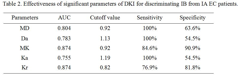
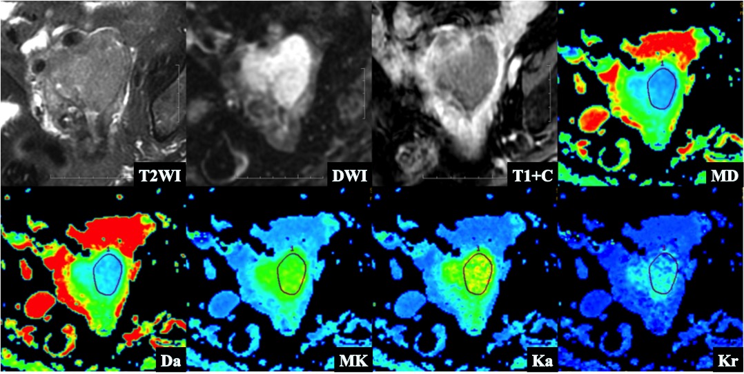
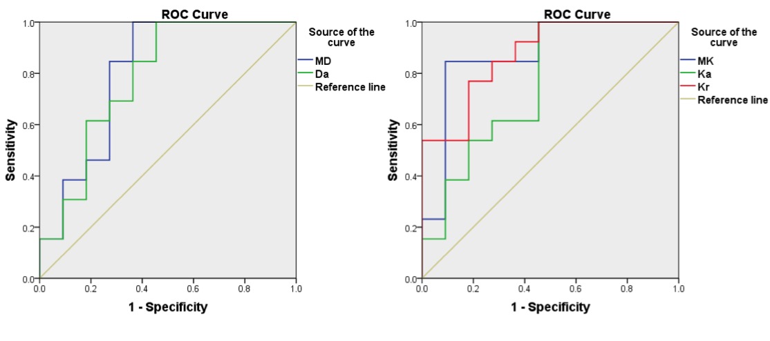
Twenty four pathologically proved patients, classified into two groups as follows: 13 in stage IA and 11 in stage IB, were enrolled, they were scanned by 1.5T MR unit with DKI sequence before treatment. Fractional anisotropy (FA), mean diffusivity (MD), axial diffusivity (Da), radial diffusivity (Dr), fractional anisotropy of kurtosis (FAk), mean kurtosis (MK), axial kurtosis (Ka) and radial kurtosis (Kr) were derived from DKI. The freehand ROI was drawn three times to take average, along the border of the low signal comprising the tumor to cover the maximum axial tumor parenchyma area. These parameters were compared in different groups. ROC analysis was performed to evaluate the diagnostic performance and to discover the corresponding threshold.Results
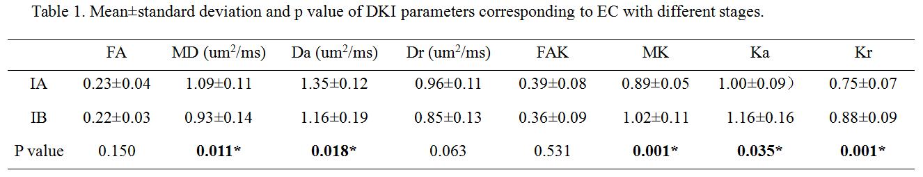
MD and Da values of stage IB group were significantly lower than that of stage IA group[( 0.93 ± 0.14 )um2/ms VS ( 1.09 ± 0.11 )um2/ms, P=0.011; ( 1.16 ± 0.19 )um2/ms VS ( 1.35 ± 0.1 )um2/ms, P=0.018], while MK, Ka and Kr values were significantly higher[( 1.02 ± 0.11 ) VS ( 0.89 ± 0.05 ), P=0.001; ( 1.16 ± 0.16 ) VS ( 1.00 ± 0.09 ), P=0.035; ( 0.88 ± 0.09 ) VS ( 0.75 ± 0.07 ), P=0.001]. The rest parameters were not found to exhibit significant difference between two groups. AUC was 0.874 for MK with sensitivity of 84.6% and specificity of 90.9% when the cutoff value was 0.92.Discussion
Diffusion kurtosis imaging (DKI), an extension of DWI, based on a non-Gaussian diffusion model better reckons for restricted water diffusion within the complex microstructure of the biological tissues [3]. We performed MD and Da values to be lower, MK, Ka and Kr values to be significantly higher, in lesions staged IB. A possible explanation is that there is more marked variation in cell size and shape within lesions staged IB in comparison with lesions staged IA [4].Conclusion
The pilot study demonstrated that DKI sequence can be used to distinguish stage IA and IB of EC, and has potential value to be a non-enhancement quantitative index for staging EC.Acknowledgements
No acknowledgement found.References
[1] Beddy P, O'Neill A C, Yamamoto A K, et al. FIGO staging system for endometrial cancer: Added benefits of MR imaging[J]. Radiographics, 2012, 32(1):241.
[2] Beddy P,Moyle P,Kataoka M,et al. Evaluation of depth of myometrial invasion and overall staging in endometrial cancer: Comparison of diffusion- weighted and dynamic contrast-enhanced MR imaging[J]. Radiology,2012,262(2):530-537.
[3] Jensen JH, Helpern JA, Ramani A, et al. Diffusional kurtosis imaging: The quantification of non-gaussian water diffusion by means of magnetic resonance imaging[J]. Magnetic Resonance in Medicine, 2005, 53(6): 1432-1440.
[4] Yu J, Huang D Y, Li Y, et al. Correlation of standard diffusion-weighted imaging and diffusion kurtosis imaging with distant metastases of rectal carcinoma.[J]. Journal of Magnetic Resonance Imaging, 2016, 44(1): 221-229.
Figures